


In a study published in the Journal of Microbiology & Biology Education on November 8, 2024, a team led by Professor Masaharu Takemura at the Tokyo University of Science has successfully captured the viral infection process under a light microscope, creating a stunning video showcasing their results. The key to this process was a unique ‘giant’ virus known as Mimivirus. This research was co-authored by Ms. Kanako Morioka and Ms. Ayumi Fujieda at Tokyo’s Yone Production Co., Tokyo, Japan.
Mimivirus has a much larger particle size than most viruses and can actually be seen under a light microscope, making it an ideal candidate for use as an educational tool. The researchers sought to visualize how the Mimivirus infects a microbe called Acanthamoeba. It is
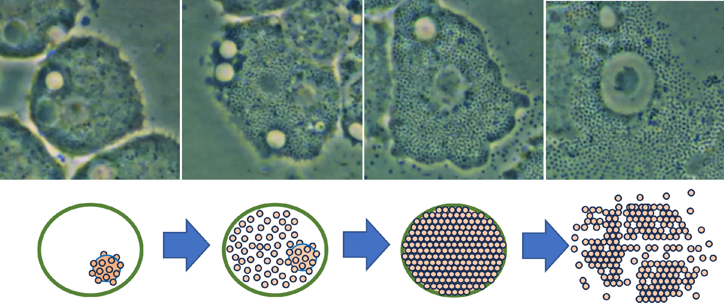
difficult to visualize amoebae under a microscope since they are constantly moving in a liquid medium; therefore, they used a modified growth medium containing a jelly-like substance called agar. This growth medium also contained viruses which infected the amoebae, and after infection, the Acanthamoeba cells moving under the agarose gel gradually slowed down.
The researchers were able to film individual cells as they were infected; indeed, we can observe all the steps of the viral infection process in their footage. While healthy Acanthamoeba cells are initially moving around, they gradually slow down and come to a stop following Mimivirus infection. As the amoeba cells stopped moving, the researchers observed the development of a ‘virion factory’ inside the amoeba cell, which produced more ‘virions’ or viral particles. The infected cell ultimately dies as its membrane ruptures.
Prof. Takemura highlights the study’s innovation, saying, “For the first time in the world, we have succeeded in continuously visualizing the events that are believed to occur in viral infection over a long period of time—such as the proliferation of the virus, its release from
cells, and the death of cells during the process.”
The film showing how a single Acanthamoeba cell is infected by Mimivirus was then screened in a biology classroom at the Tokyo University of Science and garnered positive reactions. The researchers observed that the movie influenced the perception of some students regarding viruses and seems to have shifted their views towards more scientific and biological perspectives.
This study also ensures that there is no violation of biological safety guidelines since the Acanthamoeba cells and viruses are grown in an appropriately equipped laboratory. The students in the classroom do not actually handle any of the equipment; the focus is only on
screening the filmed video in a classroom setting.
Prof. Takemura is confident that this film will be a valuable tool for teaching biology, explaining that, “It enhances students’ understanding of virus proliferation mechanisms and highlights the biological significance of viruses, their impact on host cell fate, and their role in ecosystems.”


.jpg)