Russian and Italian scientists discover new substance that can kill cancer cells

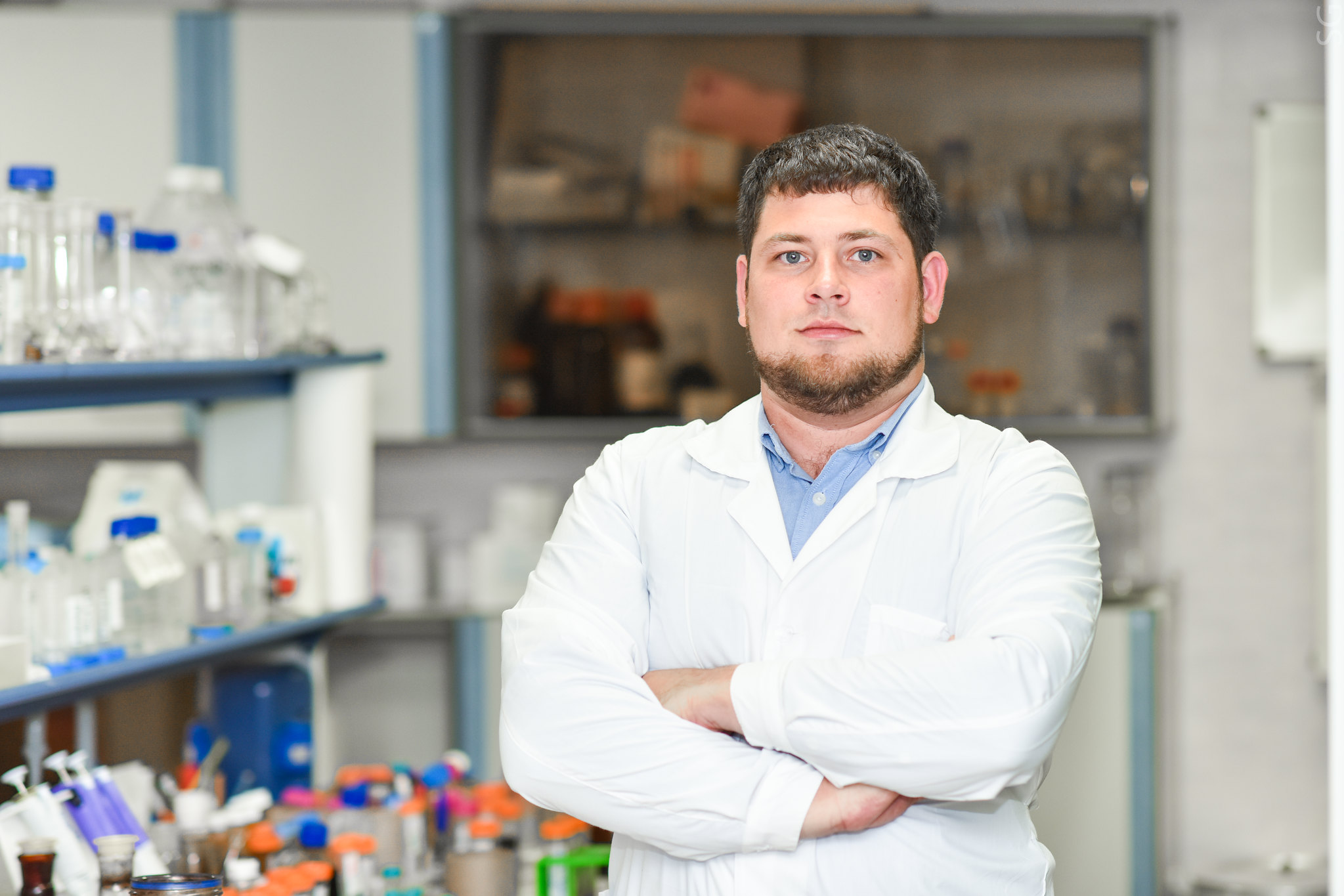
Specialists of the Center of Medical Chemistry of Togliatti State University in collaboration with colleagues from Saint Petersburg State University and University of Florence (Università degli Studi di Firenze) have found a new pro-apoptotic agent – a substance capable to suppress the growth of malignant tumours.

In March of this year, the specialists of  TSU, SPbSU and UoF conducted a joint research study that resulted in new chemical substances from the group of sulfonamides that inhibit the activity of carbonic anhydrase (CA).

Carbonic anhydrases (CAs) – is an important class of enzymes in the human body that are responsible for the regulation of various physiological processes, ensuring the constancy of the internal environment of the cell in terms of CO2 level and pH –balance. A cancer cell, unlike a normal one, has various mechanisms for survival, one of them being an increased expression of carbonic anhydrase.

In an unfavourable environment, a cancer cell begins to intensively express (synthesize) carbonic anhydrase on its surface, which “acidifies” everything around, killing healthy
cells and creating conditions for tumour growth.

In search of new carbonic anhydrase inhibitors, scientists from two countries received an unexpected result.

” This time we tried a new class of inhibitors, which should have had a slightly different
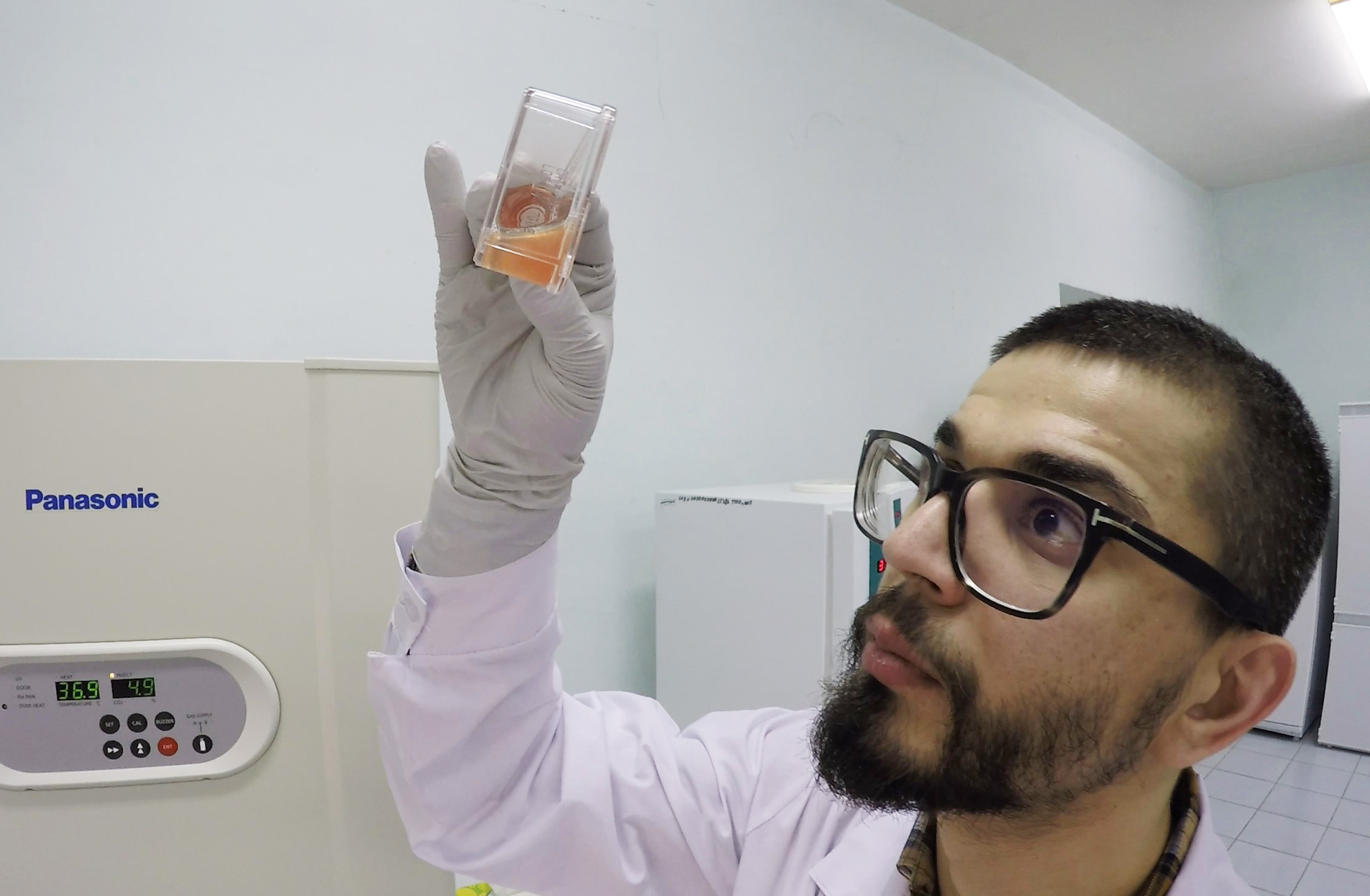
mechanism of carbonic anhydrase inactivation. Unfortunately, our substances did not work according to this mechanism, but it turned out that one of the compounds had an activity that did not correlate with the activity of carbonic anhydrase. This is how we discovered a new pro-apoptotic agent – ” Alexandr Bunev, director of the Center for Medical Chemistry, said.

Apoptosis is one of the most conservative mechanisms of cell death, which is necessary for maintaining cellular homeostasis*. In a normal cell, it is triggered in the case of some disorders or damage, while the cancer cell does everything to suppress apoptosis.

” A cancer cell does not need apoptosis, on the contrary, it acquires some resistance to this
process due to incorrect mutations and divisions. From this point of view, apoptosis inducers – chemicals that can affect also tumour cells and induce (cause) apoptosis in them – represent a fundamentally interesting mechanism of action in modern antitumor, including targeted drugs, ” Alexander Bunev explains.

Scientists conducted a series of tests to confirm that under the influence of the new compound, cancer cells entered deep apoptosis. Studies have also allowed experts to assume that the resulting substance is able to intercalate (penetrate) into Deoxyribonucleic acid (DNA).

This gives a certain failure in the division, and the cell is forced to go into apoptosis, even if it has some algorithms for bypassing it, –Alexander Bunev says.

The results of the scientists’ joint work from Togliatti, St. Petersburg and Florence are published in the European Journal of Medical Chemistry (Q1), which provides coverage to
the original research works in the main fields of medicinal chemistry.

Scientists improve contrast in noninvasive imaging of cancer cells

A Russian-German research team has come up with a new technique for magnetic resonance imaging of cancer cells. The study, published in Pharmaceutics, shows that heterologous expression of encapsulin systems from Quasibacillus thermotolerans with functional cargo proteins and iron transporter leads to increased contrast in MRI imaging of mammalian tumor cells.

Many advances in cancer treatment would come from a better understanding of tumor biology, particularly the elucidation of carcinogenesis mechanisms.

Currently, the primary method of live-cell imaging is direct labeling of cells with a probe or contrast agent before transplantation. However, any synthetic contrast agent for cell labeling has a critical drawback—it dilutes as the cells divide, which leads to loss of the signal after several cycles of divisions. In contrast, genetically encoded reporters propagate to daughter cells with each cell division. Moreover, because genetically encoded reporters rely on essential cellular processes, their signal is selective for viable cells.

The most commonly studied genetically encoded labels use an optical signal generated by either bioluminescent or fluorescent reporter protein. Although these methods have very high sensitivity, their use is limited by light scattering in biological tissues.

MRI has the advantage of deep tissue penetration with relatively high spatial resolution. Ferritin, a blood protein that contains iron, is the most studied genetically encoded agent so far. Nevertheless, ferritin’s MRI performance is severely limited by its weak magnetic properties and highly conservative structure. The latter excludes significant improvement in ferritin relaxivity by bioengineering.

“One of the most promising approaches is based on the heterologous expression of bacterial protein nanocompartments—encapsulins— in mammalian cells. Encapsulins, which are bacterial protein nanocompartments, can serve as genetically controlled labels for multimodal detection of cells. Such nanocompartments can host various guest molecules inside their lumen,” says Maxim Abakumov, head of the NUST MISIS Biomedical Nanomaterials Laboratory, senior researcher at the Medicinal Nanobiotechnology Department, N.I. Pirogov Russian National Research Medical University.

“These include, for example, fluorescent proteins or enzymes with ferroxidase activity leading to biomineralization of iron oxide inside the encapsulin nanoshell. Besides, these reporters do not suffer from dilution during cell division.”

In their experiments, a team of scientists from NUST MISIS, V. Serbsky National Medical, N.I. Pirogov Russian National Research Medical University, Technical University of Munich, Helmholtz Center Munich have implemented, for the first time, heterologous expression of encapsulin systems from Quasibacillus thermotolerans using a fluorescent reporter protein and ferroxidase in human hepatocellular carcinoma cells.

Gene expression is the process by which information from a gene is used in the synthesis of a functional gene product that enables it to produce end products, protein or non-coding RNA, and ultimately affect a phenotype, as the final effect. The researchers loaded the nanoshell with the natural ferroxidase cargo from Q. thermotolerans and a synthetic fluorescent cargo protein derived from mScarlet-I.

The successful expression of self-assembled encapsulin nano compartments with functional cargo proteins was then confirmed by fluorescence microscopy and transmission electron microscopy. Also, coexpression of encapsulin nanoshells, ferroxidase cargo, and iron transporter led to an increase in contrast in magnetic resonance imaging of cancer cells. The encapsulin cargo system from Q. thermotolerans may be suitable for multimodal imaging of cancer cells, the researhers believe.

HKBU and Cornell University jointly develop a novel targeted therapy for breast cancer

Researchers from Hong Kong Baptist University (HKBU), in collaboration with Cornell University, have developed a novel targeted therapy for triple-negative breast cancer (TNBC) that uses a specially-designed nano-carrier to deliver the Chinese medicine compound gambogic acid (GA).

The invention enhances the anti-cancer effect of GA and reduces its damage to off-target organs. The invention has the potential to become a more effective therapeutic option for TNBC.

The study was supported by the Vincent and Lily Woo Foundation, and the research findings have been published in the international medical journal Frontiers in Oncology.

GA as a breast cancer treatment and its limitations

TNBC accounts for 10-24% of all breast cancer cases and it also grows and spreads faster than other types of breast cancer. There are limited treatment options for TNBC and it has a high risk of recurrence and metastasis. In the advanced stage of the disease, the five-year relative survival rate is only about 12%.

GA is a herbal compound isolated from a dry, brownish resin called gamboge, which is derived from Garcinia hanburyi, a plant with a long history of medicinal use in Southeast Asia.

Previous studies have shown that GA can inhibit the growth of cancer cells. However, its clinical application is limited by the fact that it is rapidly eliminated from the circulation system and has poor water solubility, which makes it difficult for GA to reach the cancer cells. Furthermore, high dosages of GA can cause damage to off-target organs due to its toxicity.

Nano-carrier increases treatment efficacy of GA

In the search for a more effective treatment protocol for TNBC when compared to existing options, Professor Bian Zhaoxiang, Director and Tsang Shiu Tim Endowed Professor in Chinese Medicine Clinical Studies of the Clinical Division of HKBU’s School of Chinese Medicine (SCM) and Dr Kwan Hiu-yee, Assistant Professor of the Teaching and Research Division of SCM, together with the research team of Professor Chu Chih-Chang at Cornell University, designed a novel nano-carrier to enhance GA’s efficacy as a TNBC treatment and reduce its off-target toxicity.

The researchers made a bio-degradable nano-carrier out of polyester urea urethane (PEUU), and they decorated it with folate (also known as vitamin B9) and arginine (an amino acid). Folate receptors are highly expressed in TNBC cells, and they can serve as a target for therapy. Arginine is a positively charged amino acid, and it can attract the nano-carrier to the negatively charged tumour surface. These features enable the nano-carrier to target and deliver GA effectively to TNBC cells.

Treatment efficacy tested in mice

The research team tested the efficacy of the GA-loaded nano-carrier as a TNBC treatment in a series of mouse experiments. Two groups of mice with TNBC were treated with the same dosage of GA, one in the form of the GA-loaded nano-carrier, and the other in the form of free GA. After 17 days of treatment, the average reduction in tumour weight of the GA-loaded nano-carrier group was 67.6% higher than the free GA group. The results showed that the GA-loaded nano-carrier is more effective at shrinking the tumours than the free GA.

In addition, the group treated with the GA-loaded nano-carrier had 0.23 μg/mL of GA in their tumours two hours after injection, and the tumour GA concentration of the GA-loaded nano-carrier group was three times of the free GA group, showing that GA is being delivered to TNBC cells more effectively with the nano-carrier. Also, the concentration of GA in the plasma of the GA-loaded nano-carrier group two hour after injection was nearly three times of the free GA group, showing that the GA carried by the nano-carrier stays in the circulation system for longer.

Reduced off-target damage to other organs 

Furthermore, when compared with free GA, the GA delivered by the nano-carrier caused less damage to the off-target organs of the mice including their hearts, livers and lungs. It also caused minimal damage to their kidneys and spleens as relatively low levels of GA were detected in these two organs.

“As demonstrated in our study, the novel nano-carrier for GA offers many benefits when it comes to treating TNBC,” said Dr Kwan Hiu-yee.

“The application of nanotechnology in this study modernises the delivery of Chinese medicine, thereby enhancing its therapeutic efficacy. We believe that our nano-carriers have great clinical potential to treat TNBC and other types of cancer,” said Professor Bian Zhaoxiang.

Novel “Hydrogel” Carriers for Anti-Cancer Drugs Offer New Hope for Cancer Treatment

Hydrogels are often used as drug delivery systems, but to be effective carriers for anti-cancer drugs, they need to be responsive to varied stimuli in the tumour microenvironment. Now, scientists from Japan have developed novel hydrogels to effectively deliver drugs to tumour sites in response to temperature and pH changes in the tumour microenvironment.

These multi-stimuli-responsive hydrogels can eliminate remnant cancer cells following tumour excision through controlled drug release, offering hope for effective cancer treatment.

A team of scientists, led by Professor Akihiko Kikuchi from Tokyo University of Science, reports the production of unique degradable hydrogels that respond to changes under multiple conditions in “reducing” environments mimicking the microenvironment of tumors.

As Prof. Kikuchi observes, “In order to prepare degradable hydrogels that can release drugs in response to changes in the tumor microenvironment, we prepared hydrogels that respond to temperature, pH, and reducing environment, and analyzed their properties.”

In their study published in the Journal of Controlled Release, Prof. Kikuchi—along with his colleagues from Tokyo University of Science, Dr. Syuuhei Komatsu, Ms. Moeno Tago, and Ms. Yu Ando, and his collaborator on the study, Prof. Taka-Aki Asoh from Osaka University—details the steps of designing these novel hydrogels from the synthetic polymer poly(ethylene glycol) diglycidyl ether and the sulfur-containing organic compound cystamine. In response to low temperatures, these hydrogels swell up while they shrink at the physiological temperature.

Additionally, the hydrogels respond to pH changes by virtue of possessing tertiary amino groups. It must be noted here that the pH of the tumour microenvironment fluctuates between 5.5 and 6.5 owing to glycolysis in the tumour cells. Under the reducing conditions of this environment, the hydrogels degrade because of the breakage of disulfide bonds and change into low molecular-weight water-soluble oligomers that are easily excreted from the body.

To further test their drug release properties, the scientists loaded these hydrogels with specific proteins by exploiting their temperature-dependent swelling-deswelling behavior and tested the controlled release of drugs under acidic or reducing conditions.

It was found that the amount of drug loaded onto these hydrogels could be controlled by changing the mesh size of the hydrogel polymer network by changing temperature, suggesting the possibility of customizing these DDSs for specific drug delivery. Besides, the hydrogel network structure and electrostatic interactions in the network ensured that the proteins were preserved intact until delivery, unaffected by the swelling and shrinking of the hydrogels with pH changes in the surrounding environment.

The scientists found that the loaded protein drugs were completely released only under reducing conditions.

Using these hydrogels and the traceability that they provide, doctors may soon be able to design “customized” hydrogels that are specific to patients, giving personalized medicine a big boost. In addition to that, this new DDS provides a way to kill cancer cells that are left behind after surgery.

As Prof. Kikuchi states, “The implantation of this material in the affected area after cancer resection may eliminate residual cancer cells, making it a more powerful therapeutic tool”.

As cancer tightens its vice grip around the world, treatment options need to be varied and upgraded for customized and effective therapy. This unique and simple design technique to produce multi-stimuli-responsive hydrogels for effective drug delivery to target tumour sites may just be one among several such promising techniques to mount an answer to the challenge cancer poses to humanity.