


Solid oxide fuel cells (SOFCs) are a promising avenue to meet global demands for clean energy. They can produce electricity through environmentally friendly electrochemical reactions. However, existing SOFCs operate at high temperatures, which lowers their efficiency. Now, researchers from Japan have developed a novel perovskite-based anode material for SOFCs that exhibits mixed hole–proton conduction at medium-range temperatures. Their findings will help establish more efficient energy technologies, leading the way to more sustainable societies.
Amidst the ongoing energy and climate crises, the stakes have never been higher. We are pressed for time to find better ways of producing clean energy to replace fossil fuels. Thus far, fuel cells appear to be one of the most promising research directions. These electrochemical devices can produce electricity directly from chemical reactions, which can be tailored to be environmentally friendly in terms of their reactants and outputs.
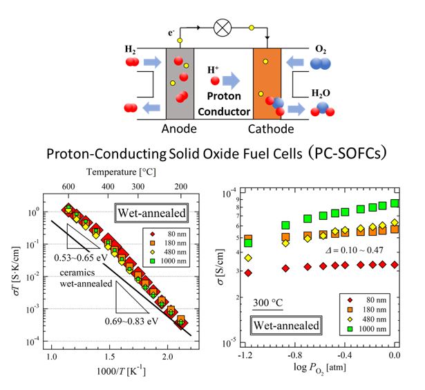
Various types of fuel cells exist, but solid oxide fuel cells (SOFCs) have attracted special attention from researchers. By operating without the need for a liquid electrolyte, they offer higher safety and are often easier to manufacture. Unfortunately, one of their main drawbacks is their high operating temperature. Conventional SOFCs need to be at over 700 °C to work properly, which limits their applicability, reduces their efficiency and power output, and often compromises their durability. Thus, proton-conducting SOFCs (PC-SOFCs), which can operate within a lower temperature range, are being investigated as a promising alternative.
Against this backdrop, a research team including Professor Tohru Higuchi from Tokyo University of Science has achieved a breakthrough in PC-SOFCs by developing a novel hole–proton mixed-conductor material. Their findings, which have been published in the Journal of the Physical Society of Japan on June 18, 2024, could pave the way for important technological advancements in energy technologies.
The material in question is a perovskite-type oxide ceramic with the formula BaCe0.4Pr0.4Y0.2O3−δ (BCPY). These particular dopants, namely Pr and Y ions, were selected based on previous works by members of the research team. They observed that BaCe0.9Y0.1O3−δ and BaPrO3−δ exhibited proton and hole (a type of positive charge carrier) conduction, respectively. Thus, they theorized that co-doping with both Pr and Y might lead to high proton–hole mixed conductivity.
Such a material could be used in the anode electrode of PC-SOFCs, as Prof. Higuchi explains: “The Pt metal electrode used in other fuel cells has issues, such as a large drop in power output because electrochemical reactions occur only at the three-phase interface where the fuel gas/electrode/electrolyte intersect. To solve this issue, a dense membrane with mixed conduction could be useful for improving the performance of PC-SOFC by expanding the electrochemical reaction area.”
Using a sputtering technique, the researchers produced thin films of BCPY and carefully analyzed its conduction properties, seeking to find evidence of mixed proton–hole conduction. To this end, they established a quantitative evaluation method to determine oxygen vacancies using X-ray absorption spectroscopy and defect chemistry analysis. Through these and several additional experiments, including synchrotron radiation photoelectron spectroscopy for electronic band structure analysis, they found substantial evidence that mixed hole–proton conductivity can occur on the surface of the proposed electrode material.
Notably, BCPY electrodes exhibited a high conductivity of over 10−2 S.K/cm at 300 °C, which outlines a bright future not only for PC-SOFCs, but for other technologies as well. “If we can further confirm that BCPY thin films do enable hole–proton mixed conductivity, BCPY may become a novel oxide material for not only PC-SOFC anode electrode membranes but also electric-double-layer-transistors,” highlights Prof. Higuchi. To clarify, this transistor technology can address the scalability and miniaturization problems of conventional transistors, which will be crucial to developing artificial intelligence systems and increasing the computational capacity of personal electronic devices.
In any case, this study sheds some much-needed light on new electrode materials for PC-SOFCs. With further advances in this exciting field, electrochemical energy generation could eventually enable us to power up our homes and cars with cleaner electricity, paving the way to more sustainable societies.
.jpg)

.jpg)