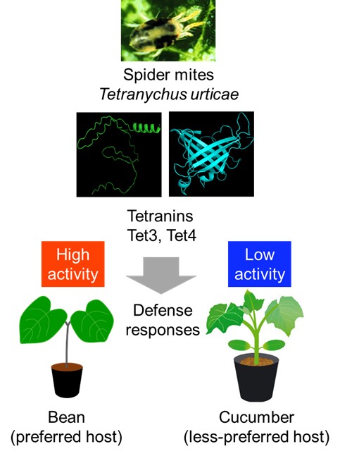
Breakthrough in Z-Alkene synthesis: Scientists develop efficient, sustainable method

The photoisomerization of E-to-Z alkenes has many applications in diverse fields, including organic chemistry, polymer chemistry, and medicinal chemistry. In a new study, researchers from Japan developed a new closed-loop method for photoisomerization of E-to-Z alkenes using a recycling photoreactor. This innovative method utilizes the high-performance liquid chromatography method to recycle the samples, thereby improving efficiency. This eco-friendly method can lead to the sustainable development of various chemicals, including pharmaceuticals.

Z-alkenes are organic compounds with a double bond between two carbon atoms and two substituents attached to the carbon atoms on the same side of the double bond. They are ubiquitous structural components of organic compounds in chemistry and biology. It is well known that many of the Z-alkenes cannot be prepared through conventional methods involving thermodynamic methods while photoisomerization can offer good yields. Photoisomerization is a process in which the structural arrangement of an isomer of a molecule is changed to another isomer by absorption of light. The photoisomerization of E-alkenes to produce Z-alkenes has many applications in the fields of organic chemistry, polymer chemistry, and medicinal chemistry.

Many studies have explored different methods for photoisomerization of E-to-Z alkenes. A notable approach involved a continuous-flow system, in which the photosensitizer was immobilized in an ionic liquid and continuously recycled via a simple phase separation process. Photosensitizers are materials that enhance the rate of photoisomerization reactions by absorbing light energy and transferring it to the reactant molecule. However, current methods based on the use of ionic liquids are time-consuming and difficult to apply to recycling high-performance liquid chromatography (HPLC) technology, which enables the recycling of samples and therefore can enhance the efficiency of photoisomerization.

Inspired by these findings, in a new study, a team of researchers from Japan, led by Professor Hideyo Takahashi from the Faculty of Pharmaceutical Sciences at Tokyo University of Science, explored the photoisomerization of E-cinnamamides to Z-cinnamamides using a recycling photoreactor coupled with an HPLC system. The study, published online in The Journal of Organic Chemistry on June 05, 2024, included contributions from Ms. Mayuko Suga, Ms. Saki Fukushima, and Assistant Professor Dr. Kayo Nakamura, also from Tokyo University of Science.

The team had previously developed a recycling photoreactor based on the deracemization concept. Prof. Takahashi explains, “Our closed-loop recycling photoreactor was initially used to convert a racemate, a mixture of left and right-handed enantiomers of a chiral molecule, into the pure desired enantiomer. It consists of a photocatalyst immobilized on a resin, which converts an undesired enantiomer into a racemate, and an HPLC column which separates the desired enantiomer. In this study, we adapted this method to convert E-cinnamamides to their Z-isomers.”
To employ this method for E-to-Z photoisomerization of alkenes, a photosensitizer that promotes rapid photoisomerization is required. To this end, the researchers screened several commercially available photosensitizers and identified thioxanthone as the best candidate. Next, they investigated its immobilization. Thioxanthone, with functional amide groups as linkages, was immobilized on a modified silica gel. This immobilization not only prevented the leakage of photosensitizer in the solid phase but also enhanced the catalytic activity compared to the parent soluble thioxanthone. This enhancement was particularly interesting, as solid-phase reactions are typically slower than liquid-phase reactions.

This superior catalytic activity was attributed to the introduction of suitable functional groups. The researchers, therefore, evaluated the catalytic activity of various photosensitizers with different functional groups by comparing the total amount of light required for promoting photoisomerization. With the optimal photosensitizer identified, they conducted the photoisomerization reactions in the recycling photoreactor, yielding the desired Z-alkenes in good yields after 4–10 cycles.

“This recycling photoreactor shows promise as an efficient alternative system to produce Z-alkenes,” remarks Prof. Takahashi. “Due to the continuous closed-loop recycling of the samples, it represents an environmentally friendly and sustainable method.”

This innovative method can lead to more eco-friendly development of Z-alkenes, and therefore pharmaceuticals, paving the way towards a sustainable future.

National Taiwan University expands student exchange with Kyushu U and UIUC, by establishing joint center

On February 19, President Tatsurou Ishibashi of Kyushu University, Japan, and President Robert Jones of the University of Illinois at Urbana-Champaign (UIUC), USA, led delegations to visit National Taiwan University (NTU). Together with NTU President Wen-Chang Chen, they signed a Memorandum of Understanding (MOU) for the NTU-Kyushu-UIUC Joint Center and unveiled the center’s plaque. This collaboration aims to expand inter-university exchanges to a trilateral framework, systematically promoting faculty and student exchanges, as well as research collaborations in fields such as medicine, engineering, agriculture, and humanities and social sciences. Deputy Minister of Education Liu Meng-chi also attended the luncheon to congratulate the three universities on this new milestone in international cooperation.

Both Kyushu University and UIUC are key partner institutions of NTU. The exchange of students, dual-degree programs, and other collaborative efforts between these universities have been steadily developing. Numerous bilateral meetings have further connected their engineering, electrical engineering, agricultural, and social sciences departments. Additionally, Kyushu University and UIUC established a key sister school relationship in 2019.

One-step synthesis of pharmaceutical building blocks: New method for anti-Michael reaction

The anti-Michael addition reaction, which involves nucleophilic addition reactions to the α-position of α,β-unsaturated carbonyl compounds, has been difficult to achieve so far. In a new study, researchers from Japan have developed a new method for successful anti-Michael addition reaction of α-unsaturated carbonyl compounds, which are commonly used in pharmaceuticals. This reaction is expected to be used as a one-step synthesis method with 100% atomic efficiency for α-unsaturated carbonyl compounds.

In 1887, chemist Sir Arthur Michael reported a nucleophilic addition reaction to the β-position of α,β- unsaturated carbonyl compounds. These reactions, named Michael addition reactions, have been extensively studied to date. In contrast, the anti-Michael addition reaction, referring to the nucleophilic addition reaction to the α-position, has been difficult to achieve. This is due to the higher electrophilicity of the β-position compared to the α-position. Previous attempts to overcome these difficulties have involved two main methods. The first is restricting the addition position via intramolecular reactions, while the second method involves introducing a strong-electron withdrawing group at the β-position. However, these methods are not ideal for synthesizing complex molecules via the anti-Michael reaction.

In a new study, a global team of researchers, led by Professor Takanori Matsuda and including Mr. Ryota Moro, both from the Department of Applied Chemistry at Tokyo University of Science, Japan, as well as including Assistant Professor Hirotsugu Suzuki from the Tenure-Track Program for Innovative Research at the University of Fukui, Japan, successfully achieved palladium-catalyzed anti-Michael addition reaction of acrylamides. This represents the first example of an anti-Michael-type addition reaction. “We found that the presence of a catalytic amount of palladium(II) trifluoroacetate Pd(TFA)2 is capable of facilitating the anti-Michael addition of indole to acrylamide with an aminoquinoline group as a directing group, producing the addition product in high-yield,” explains Prof. Matsuda.

Their study was made available online on May 14, 2024, and published in Volume 146, Issue 20 of the Journal of the American Chemical Society on May 22, 2024.

The team reasoned introducing a directing group into an α,β-unsaturated carbonyl compound could facilitate an anti-Michael type addition reaction by stabilizing the reaction intermediate. To test this, the researchers first used an acrylamide having an aminoquinoline-directing group, and a nucleophile, 1-methylindole, as model substrates to investigate the anti-Michael type addition reaction in the presence of the palladium catalyst. This reaction produced the desired product with a 90% yield. At a reaction scale of two millimoles, there was no yield loss, signifying the practicality of the reaction.

This reaction was also carried out with β-substituted cinnamamide derivatives and crotonamide derivatives with an alkyl group. Moreover, the reaction proceeded smoothly with a wide range of nucleophiles, including many indoles, heterocyclic compounds such as pyrroles and thiophenes, and electron-rich aromatic compounds. Additionally, the aminoquinoline-directing group used in this reaction can be converted to carboxylic acids and other amides, signifying the usefulness of the reaction.

The researchers also investigated the mechanism for this reaction through labeling experiments. They found that initially, the acrylamide coordinates to Pd(TFA)2 to form a five-membered ring palladacyle intermediate. The reaction then proceeds with the nucleophilic attack by indole on the intermediate, producing alkylpalladium species. Finally, an acid removes palladium and regenerates Pd(TFA)2, producing the desired α-substituted carbonyl compound.

Highlighting the potential applications of this study, Dr. Suzuki says, “The anti-Michael type addition is expected to become an ideal one-step reaction with 100% atomic efficiency for the synthesis of α-substituted carbonyl compounds, which are often used in pharmaceuticals. Our method will enable the widespread application of this reaction.”

Overall, this novel method can lead to efficient and sustainable synthesis of α-substituted carbonyl compounds and consequently pharmaceuticals, among other organic compounds.

New insights into antihistamine binding could lead to more effective treatments

Doxepin is an antihistaminic, antidepressant, and sleeping aid that has two geometric isomers—molecules with equal chemical formulas but different 3D arrangements. While its Z-isomer is known to be more effective than its E-isomer, the precise nature of its binding to the histamine H1 receptor remained elusive. Now, in a recent study, researchers from Japan thoroughly addressed this knowledge gap through an innovative experimental protocol, paving the way to next-generation antihistamines with fewer side effects.

Even if two molecules have the exact same chemical formula and the same number and types of bonds, their three-dimensional arrangements can still be different. While some people might mistakenly disregard this as a minor detail, even simple changes in the position or orientation of a functional group can dramatically affect the biological properties of a molecule, sometimes rendering an otherwise benign substance into a highly toxic one. Thus, the study of such possible molecular variants, called ‘geometric isomers,’ is essential in the field of drug development.

Doxepin stands out as a notable example of a drug that is commercialized as a mixture of two geometric isomers, namely the E- and Z-isomers. Both doxepin isomers bind to histamine H1 receptor (H1R), which is expressed throughout the central nervous system, smooth muscle cells, and vascular endothelial cells. Besides its use as an antihistaminic drug, doxepin is also typically used as an antidepressant and sleeping aid. While biological tests in animals have shown that the Z-isomer is more effective than the E-isomer, the differences in affinity to H1R between the E- and Z-isomers are unknown. Moreover, the specifics of how these compounds actually bind to H1R remain elusive.

Against this backdrop, a research team from Tokyo University of Science, Japan, set out to clarify the finer details of the interactions between doxepin isomers and H1R. Their latest paper, which was published on June 25, 2024, in the Journal of Molecular Recognition, was co-authored by Professor Mitsunori Shiroishi, Mr. Hiroto Kaneko, and Associate Professor Tadashi Ando, among others. This study is a follow-up to past work done by Prof. Shiroishi and colleagues. “We previously revealed the crystal structure of the complex formed by H1R and doxepin, but we were unable to determine which isomer was bound,” he explains, “We then came up with a method to determine the binding affinity of the isomers, and thus carried out this study.”

To achieve this challenging goal, the researchers first produced a customized yeast expression vector by strategically inserting the H1R gene into it. This vector was used to modify yeast cultures so that they produce H1R. After retrieving the membranes from these cells, they applied a solution containing commercial doxepin, producing H1R-doxepin complexes. Following extraction and purification of these complexes, they removed any excess (unbound) doxepin. Finally, by denaturing the H1R receptors, they could free the bound doxepin molecules and measure their numbers in a high-performance liquid chromatography setup.

Using this protocol, the researchers could accurately quantify the amount of each isomer that was bound to the extracted receptors, which is directly tied to their relative binding affinity. They found that the affinity to H1R of the Z-isomer was over five times higher than that of the E-isomer.

The team then delved deeper into the nature of how doxepin isomers bind to H1R. Through experiments on a mutant variant of H1R coupled with molecular dynamics simulations, they revealed that the Thr112 side chain in the ligand-binding pocket of H1R creates a chemical environment that enhances selectivity for the Z-isomer.

Taken together, the findings of this study shed light on how a widely used small molecule drug interacts with an important cellular receptor. “Our efforts could serve as the basis for designing next-generation antihistamines that are more effective and have fewer side effects,” highlights Prof. Shiroishi, “Worth noting, this newfound knowledge will be useful for designing compounds that bind not only to H1R, but also other disease-relevant target proteins.”

The rational design of future drugs, aided and validated by computational techniques like molecular dynamics simulations, could usher in a new era in medicine. More specifically, by understanding the binding properties of isomers in detail, many small-molecule drugs could be made more effective, safer, and better suited for targeted therapies.

Let us hope this vision of the future becomes a reality soon!

Towards Cleaner Energy: Breakthrough in Anode Electrode Materials for Proton Conducting Solid Oxide Fuel Cells Operating at Medium Temperature

Solid oxide fuel cells (SOFCs) are a promising avenue to meet global demands for clean energy. They can produce electricity through environmentally friendly electrochemical reactions. However, existing SOFCs operate at high temperatures, which lowers their efficiency. Now, researchers from Japan have developed a novel perovskite-based anode material for SOFCs that exhibits mixed hole–proton conduction at medium-range temperatures. Their findings will help establish more efficient energy technologies, leading the way to more sustainable societies.

Amidst the ongoing energy and climate crises, the stakes have never been higher. We are pressed for time to find better ways of producing clean energy to replace fossil fuels. Thus far, fuel cells appear to be one of the most promising research directions. These electrochemical devices can produce electricity directly from chemical reactions, which can be tailored to be environmentally friendly in terms of their reactants and outputs.

Various types of fuel cells exist, but solid oxide fuel cells (SOFCs) have attracted special attention from researchers. By operating without the need for a liquid electrolyte, they offer higher safety and are often easier to manufacture. Unfortunately, one of their main drawbacks is their high operating temperature. Conventional SOFCs need to be at over 700 °C to work properly, which limits their applicability, reduces their efficiency and power output, and often compromises their durability. Thus, proton-conducting SOFCs (PC-SOFCs), which can operate within a lower temperature range, are being investigated as a promising alternative.

Against this backdrop, a research team including Professor Tohru Higuchi from Tokyo University of Science has achieved a breakthrough in PC-SOFCs by developing a novel hole–proton mixed-conductor material. Their findings, which have been published in the Journal of the Physical Society of Japan on June 18, 2024, could pave the way for important technological advancements in energy technologies.

The material in question is a perovskite-type oxide ceramic with the formula BaCe0.4Pr0.4Y0.2O3−δ (BCPY). These particular dopants, namely Pr and Y ions, were selected based on previous works by members of the research team. They observed that BaCe0.9Y0.1O3−δ and BaPrO3−δ exhibited proton and hole (a type of positive charge carrier) conduction, respectively. Thus, they theorized that co-doping with both Pr and Y might lead to high proton–hole mixed conductivity.

Such a material could be used in the anode electrode of PC-SOFCs, as Prof. Higuchi explains: “The Pt metal electrode used in other fuel cells has issues, such as a large drop in power output because electrochemical reactions occur only at the three-phase interface where the fuel gas/electrode/electrolyte intersect. To solve this issue, a dense membrane with mixed conduction could be useful for improving the performance of PC-SOFC by expanding the electrochemical reaction area.”

Using a sputtering technique, the researchers produced thin films of BCPY and carefully analyzed its conduction properties, seeking to find evidence of mixed proton–hole conduction. To this end, they established a quantitative evaluation method to determine oxygen vacancies using X-ray absorption spectroscopy and defect chemistry analysis. Through these and several additional experiments, including synchrotron radiation photoelectron spectroscopy for electronic band structure analysis, they found substantial evidence that mixed hole–proton conductivity can occur on the surface of the proposed electrode material.

Notably, BCPY electrodes exhibited a high conductivity of over 10−2 S.K/cm at 300 °C, which outlines a bright future not only for PC-SOFCs, but for other technologies as well. “If we can further confirm that BCPY thin films do enable hole–proton mixed conductivity, BCPY may become a novel oxide material for not only PC-SOFC anode electrode membranes but also electric-double-layer-transistors,” highlights Prof. Higuchi. To clarify, this transistor technology can address the scalability and miniaturization problems of conventional transistors, which will be crucial to developing artificial intelligence systems and increasing the computational capacity of personal electronic devices.

In any case, this study sheds some much-needed light on new electrode materials for PC-SOFCs. With further advances in this exciting field, electrochemical energy generation could eventually enable us to power up our homes and cars with cleaner electricity, paving the way to more sustainable societies.

Towards Next-Gen Functional Materials: Direct Observation of Electron Transfer in Solids

Nanoscale electron transfer (ET) in solids is fundamental to the development of multifunctional materials. However, ET in solids is not yet clearly understood. Now, researchers from Japan achieved a direct observation of solid-state ET through X-ray crystal analysis by fabricating a novel double-walled non-covalent crystalline nanotube, which can absorb electron donor molecules and maintain its crystalline structure during ET. This innovative approach can lead to the design of novel functional materials soon.

Electron transfer (ET) is a process in which an electron is transferred from one atom or molecule to another. ET is fundamental to electrochemical reactions with applications in many fields. Nanoscale ET, which involves the transfer of electrons in the range of 1–100 nanometers in solids is fundamental to the design of multifunctional materials. However, this process is not yet clearly understood.

Nanotubes, nanomaterials with unique cylindrical nanostructures, offer a variety of ET properties that can be realized through electron and hole (vacant spaces left by electrons) injections into the nanotubes, making them a suitable candidate for studying nanoscale ET. Although carbon-based nanotubes have fascinating ET properties, they are particularly difficult to control in terms of their shape and size due to extreme conditions, such as high temperatures, required for their synthesis. A viable approach for fabricating well-defined tunable nanotubes is bottom-up fabrication of non-covalent nanotubes, which sometimes result in crystalline-form nanotubes. Non-covalent nanotubes are formed through the inherent attractive interactions or non-covalent interactions between atoms, instead of the strong covalent interactions seen in carbon nanotubes. However, they are not strong enough to endure electron and hole injections, which can break their non-covalent interactions and destroy their crystalline structure.

In a recent study, a team of researchers from the Department of Applied Chemistry at Tokyo University of Science, led by Professor Junpei Yuasa and including Dr. Daiji Ogata, Mr. Shota Koide, and Mr. Hiroyuki Kishi, used a novel approach to directly observe solid-state ET. Prof. Yuasa explains, “We have developed crystalline nanotubes with a special double-walled structure. By incorporating electron donor molecules into the pores of these crystalline nanotubes through a solid-state oxidation reaction, we succeeded in directly observing the electron transfer reaction in the solid using X-ray crystal structure analysis.” Their findings were published in the journal Nature Communications on May 23, 2024.

The researchers used a novel supramolecular crystallization method, which involves oxidation-based crystallization, to fabricate zinc-based double-walled crystalline nanotubes. This double-walled structure with large windows in the nano-tube walls makes the crystal robust and flexible enough to maintain its crystalline state when subjected to ET oxidation processes. Moreover, this structure allows the crystal to absorb electron donor molecules. The researchers used ferrocene and tetrathiafulvalene as electron donor molecules, which were absorbed through the windows of the nanotube crystals. This allows electrons to be removed from the absorbed electron donors through solid-state ET oxidation reactions, resulting in the accumulation of holes in the donors inside the nanotube. Due to the robustness of the crystals, the researchers were able to observe this ET oxidation process using X-ray crystal structure analysis directly, uncovering key insights.

This novel approach is highly valuable for direct observation of ET in solid nanomaterials. Highlighting the potential applications of this study, Prof. Yuasa says, “Understanding ET can lead to the development of novel functional materials, which in turn can lead to the design of more efficient semiconductors, transistors, and other electronic devices. Optoelectronic devices, such as solar cells, rely heavily on ET. Hence, direct observation of ET can help improve these devices’ performance. Additionally, this approach can lead to advancements in energy storage, nanotechnology, and materials science research.”

Overall, this study is a striking example of direct observation of solid-state ET, which can be expanded to observe ET and related phenomena in other nanomaterials.


Fighting Fat and Inflammation: Scientists Develop Powerful New Compounds

Obesity and inflammatory diseases are increasing in prevalence and contribute to the growing burden of lifestyle disorders such as diabetes and hypertension. There is a lack of naturally derived alternatives to tackle these issues. Researchers from Tokyo University of Science have synthesized novel amino acid derivatives of menthol and studied its properties. The menthyl esters showed exceptional anti-inflammatory and anti-obesity activities during preclinical studies and can be developed as therapeutic compounds with further research.

Modified derivatives of natural products have led to significant therapeutic advances and commercial success in recent times. Menthol is a naturally occurring cyclic monoterpene alcohol found in various plants, particularly in members of the mint family such as peppermint and spearmint. It is a common ingredient found in a wide range of confectionaries, chewing gums and oral care products. Interestingly, menthol also has high medicinal value due to its analgesic, anti-inflammatory, and anti-cancer effects.

In a recent study, a team of researchers led by Professor Gen-ichiro Arimura from the Department of Biological Science and Technology, Tokyo University of Science, Japan, developed and investigated menthyl esters of valine (MV) and isoleucine (MI), which are derived from menthol by replacing its hydroxyl group with valine and isoleucine, respectively.

Their research findings were published in the Immunology journal on May 08, 2024. Sharing the motivation behind the present work, Prof. Arimura says, “The functional components of plants that contribute to human health have always intrigued me. Discovering new molecules from natural materials inspired our research team to develop these amino acid derivatives of menthol.”

The researchers began by synthesizing menthyl esters of six amino acids characterized by less-reactive side chains. Subsequently, they assessed the properties of these esters using in vitro cell line studies. Finally, they conducted experiments in mice to explore the effects of these compounds under induced disease conditions. The exceptional anti-inflammatory profiles of MV and MI was determined by assessing the transcript levels of tumor necrosis factor-α (Tnf) in stimulated macrophage cells. Remarkably, both MV and MI outperformed menthol in the anti-inflammatory assay. RNA sequencing analysis revealed that 18 genes involved in inflammatory and immune responses were effectively suppressed.

Elated with their findings, the researchers went a step further and investigated the mechanism of action of the menthyl esters. They discovered that liver X receptor (LXR) – an intracellular nuclear receptor, had an important role in the anti-inflammatory effects and this was independent of the cold-sensitive transient receptor TRPM8, which primarily detects menthol. Delving deeper into the LXR-dependant activation of MV and MI, they found that Scd1 gene – central to lipid metabolism was upregulated by LXR. Moreover, in mice with induced intestinal colitis, the anti-inflammatory effects were further validated with suppressed transcript levels of Tnf and Il6 genes by MV or MI, in an LXR-dependent manner.

Driven by the discovery of LXR-SCD1 intracellular machinery, Prof. Arimura and his team hypothesized the menthyl esters to possess anti-obesity properties. They found that these esters inhibited adipogenesis-fat accumulation, specifically at the mitotic clonal expansion stage in 3T3-L1 adipocyte cells. During animal studies, the diet-induced obesity in mice was ameliorated and adipogenesis was suppressed.

Menthyl esters possess unique advantages compared to other anti-inflammatory or anti-obesity compounds currently being researched or used. Their specific mechanisms of action, that contribute to their dual anti-inflammatory and anti-obesity effects sets them apart from other compounds and may make them particularly effective in addressing both inflammatory conditions and metabolic disorders. They could benefit specific populations like individuals with chronic inflammatory conditions, metabolic syndrome, or obesity-related complications.

“Although this study focused on their functions and mechanisms of action in diseases modeled after inflammation and obesity, we expect that these compounds will also be effective against a wide range of lifestyle-related diseases caused by metabolic syndrome, such as diabetes and hypertension, as well as allergic symptoms,” says Prof. Arimura optimistically.

In conclusion, this study underscores the importance and value of multi-faceted molecules derived from naturally occurring substances. Future research involving these novel and superior menthyl esters may result in therapeutic compounds to tackle the ever-growing health concerns of obesity and inflammatory conditions.

Gut Bacteria Metabolite Shows Promise of Fighting Inflammatory Bowel Disease

Gut bacteria convert dietary fatty acids into useful metabolites with benefits in metabolic and inflammatory disorders. However, their immunomodulatory effects and underlying molecular mechanisms remain unclear. Now, researchers from Tokyo University of Science have uncovered a novel gut bacteria-generated, fatty acid metabolite — ‘gKetoC’ which exerts protective effects against inflammatory bowel disease. Their study sheds light on molecular targets which mediate their immunosuppressive effects against intestinal bowel inflammation and highlights their therapeutic potential.

Gut microbiota or the population of microbial inhabitants in the intestine, plays a key role in digestion and maintenance of overall health. Any disturbance in the gut microbiota can, therefore, have a systemic impact. Intestinal microbes metabolize dietary components into beneficial fatty acids (FAs), supporting metabolism and maintaining host body homeostasis. Metabolites originating from polyunsaturated fatty acids (PUFAs), influenced by gut microbes such as Lactobacillus plantarum, exhibit potent effects on inflammation and immune responses. Manipulating gut bacteria and their metabolites shows promise in treating metabolic and inflammatory disorders. However, despite advances in gut health and wellness trends, the precise mechanisms governing the immunomodulatory properties of microbe-derived metabolites remain elusive.

To bridge this gap, a team of researchers led by Professor Chiharu Nishiyama from the Tokyo University of Science conducted a series of experiments using both in vitro and in vivo mouse models to understand how bacteria-generated FAs regulate immune responses. Explaining the rationale behind their work published in Frontiers in Immunology on 30 April, 2024, Prof. Nishiyama says, “PUFAs undergo metabolic transformations such as hydroxylation and saturation by enzymes possessed by intestinal bacteria. In recent years, a variety of beneficial physiological effects have been discovered for these intestinal bacterial metabolites. In this study, we have investigated the activity of multiple FA metabolites using mouse-derived immune cells.”

To this end, the researchers used antigen-stimulated spleen cells to elicit an enhanced immune response. Subsequently, they investigated the impacts of different polyunsaturated fatty acid (PUFA) derivatives, focusing on metabolites of linoleic acid, a prevalent dietary fatty acid. Their findings revealed that KetoC, αKetoC, gKetoA, and gKetoC (enon derivatives of LA) markedly reduced the levels of interleukin 2 — a key protein that triggers the expansion of immune cells and inflammation. However, the original PUFAs in their unconverted form did not demonstrate the same immunosuppressive effects, emphasizing the critical role of bacterial conversion in activating their immunomodulatory properties. Furthermore, they observed that the enon (a functional group) FAs also suppressed prolonged T-cell proliferation and dendritic cell activation, which can lead to inflammation and autoimmune diseases. This anti-inflammatory effect was most pronounced with gKetoC. Hence, the researchers aimed to unravel the molecular mechanisms through which gKetoC exerted its immunosuppressive effects.

In addition, previous studies have shown the involvement of G protein-coupled receptors (GPCRs) and the transcription factor, NRF2, in anti-oxidant responses, which are mediated by several FA metabolites, whereas the involvement of GPCRs and NRF2 in the effects of gKetoC in dendritic cells was largely unknown. To clarify the role of these proteins in gKetoC-mediated immune responses, the researchers assessed the levels of inflammatory cytokines released from antigen-stimulated and gKetoC-treated dendritic cells. Their results suggested that gKetoC stimulated the NRF2 signaling pathway, which suppressed the production of inflammatory cytokines. Additionally, GPCR-signaling also inhibited inflammatory cytokine production in dendritic cells in an NRF2-dependent manner. This unveils a potential molecular axis governing the immunomodulatory effects of gKetoC.

To further validate their findings in vivo, the researchers used a mouse model of inflammatory bowel disease and examined immune and inflammatory responses by involving gKetoC treatment. They found that gKetoC treatment significantly reduced fibrosis-induced tissue damage in the colon, reduced colitis-induced weight loss, and improved stool scores. Furthermore, the treated mice showed decreased epithelial cell disruption and ulcers, along with reduced infiltration of immune cells and lower serum levels of inflammatory factors. Notably, the models that were deficient in NRF2 showed significant restoration of colitis-induced tissue damage following gKetoC treatment.

Overall, the present study sheds light on the potential mechanism by which gKetoC alleviates antigen-induced intestinal inflammation. Further studies are needed to understand the complex interplay between gKetoC, GPCR-signaling, and the NRF2 pathway, and uncover other potential targets of gKetoC which mediate its anti-inflammatory effects. Nevertheless, anti-inflammatory FA metabolites hold therapeutic promise in the treatment of intestinal inflammatory diseases and maintenance of gut health, as prebiotic or probiotic formulations. Sharing her concluding thoughts, Dr. Nishiyama states, “Our findings demonstrate that the compounds of dietary oils are converted into useful metabolites with anti-inflammatory effects by gut bacteria. By conducting detailed analyses at the individual, cellular, and genetic levels, we hope to understand how the food we eat daily influences the function of immune cells, and how these effects can be targeted for the prevention and mitigation of inflammatory diseases.”

In summary, while the beneficial effects of bacterial PUFA metabolites were known, this study identified gKetoC as a metabolite playing a protective role in a colitis mice model. In the long run, these findings can help improve the quality of life for patients suffering from inflammatory diseases, and augment the possibility of developing functional foods, supplements, and nutraceuticals based on these microbial metabolites. Moreover, the researchers also speculate that these developments could help in the identification and development of compounds that are capable of preventing or alleviating immune-related diseases.

Here’s wishing the team luck in their future research endeavors!

Breakthrough Rice Bran Nanoparticles Show Promise as Affordable, Targeted Anticancer Agent

Plant-derived nanoparticles have demonstrated significant anticancer effects. Researchers from Japan recently developed rice bran-derived nanoparticles (rbNPs) that efficiently suppressed cell proliferation and induced programmed cell death of only cancer cells. Furthermore, rbNPs successfully suppressed the growth of tumors in mice having aggressive adenocarcinoma in their peritoneal cavity, without any adverse effects. Given their low production costs and high efficacy, rbNPs hold great promise for developing affordable and safe anticancer agents.

Several types of conventional cancer therapies, such as radiotherapy or chemotherapy, destroy healthy cells along with cancer cells. In advanced stages of cancer, tissue loss from treatments can be substantial and even fatal. Cutting-edge cancer therapies that employ nanoparticles can specifically target cancer cells, sparing healthy tissue. Recent studies have demonstrated that plant-derived nanoparticles (pdNPs) that have therapeutic effects can be an effective alternative to traditional cancer treatments. However, no pdNPs have been approved as anticancer therapeutic agents till date.

Rice bran is a byproduct generated during rice refining process that has limited utility and low commercial value. However, it contains several compounds with anticancer properties, such as γ-oryzanol and γ-tocotrienol. To explore these therapeutic properties of rice bran, a team of researchers led by Professor Makiya Nishikawa from Tokyo University of Science (TUS) in Japan developed nanoparticles from rice bran and tested their effectiveness in mice models. Their study, published in Volume 22 of Journal of Nanobiotechnology on 16 March 2024, was co-authored by Dr. Daisuke Sasaki, Ms. Hinako Suzuki, Associate Professor Kosuke Kusamori, and Assistant Professor Shoko Itakura from TUS.

“In recent years, an increasing number of new drug modalities are being developed. At the same time, development costs associated with novel therapies have increased dramatically, contributing to the burden of medical expenses. To address this issue, we used rice bran, an industrial waste with anticancer properties, to develop nanoparticles,” explains Prof. Nishikawa.

The study evaluated the anticancer effects of rice bran-derived nanoparticles (rbNPs), which were obtained by processing and purifying a suspension of Koshihikari rice bran in water. When a cancer cell line named colon26 was treated with rbNPs, cell division was arrested and programmed cell death was induced, indicating strong anticancer effects of the nanoparticles. The observed anticancer activity of rbNPs can be attributed to γ-tocotrienol and γ-oryzanol, that are easily taken up by cancer cells resulting in cell cycle arrest and programmed cell death. Additionally, rbNPs reduced the expression of proteins, such as β-catenin (a protein associated with Wnt signaling pathway involved in cell proliferation) and cyclin D1, which are known to promote cancer recurrence and metastases. Moreover, the rbNPs reduced the expression of β-catenin only in colon26 cells without affecting the non-cancerous cells.

“A key concern in the context of pdNPs is their low pharmacological activity compared to pharmaceutical drugs. However, rbNPs exhibited higher anticancer activity than DOXIL®, a liposomal pharmaceutical formulation of doxorubicin. Additionally, doxorubicin is cytotoxic to both cancer cells and non-cancerous cells, whereas rbNPs are specifically cytotoxic to cancer cells, suggesting that rbNPs are safer than doxorubicin,” highlights Prof. Nishikawa.

To confirm the anticancer properties of rbNPs in the living body, the researchers injected rbNPs into mice having aggressive adenocarcinoma in their peritoneal cavity (enclosed by the diaphragm, abdominal muscles, and pelvis and houses organs like intestines, liver, and kidneys). They observed significant suppression of tumor growth with no adverse effects on the mice. Additionally, the rbNPs significantly inhibited metastatic growth of murine melanoma B16-BL6 cells in a lung metastasis mouse model.

Rice bran has several attributes that make it an excellent source of therapeutic pdNPs. Firstly, it is economic as compared to many other sources of pdNPs. Nearly 40% of the rice bran is discarded in Japan, providing a readily available source of raw material. Secondly, the preparation efficiency of rbNPs is higher than that of previously reported pdNPs. Besides being practical and safe as an anticancer therapeutic, the physicochemical properties of rbNPs are very stable. However, a few parameters, such as establishment of separation technologies at the pharmaceutical level, assessing production process control parameters, and evaluation of efficacy and safety in human cancer cell lines and xenograft animal models, must be investigated prior to clinical trials in humans.

In conclusion, rice bran, an agricultural waste product, is a source of therapeutic pdNPs that are affordable, effective, and safe, and has the potential to revolutionize cancer treatment in the future.

“By establishing a manufacturing method for rice bran nanoparticles with stable quality and confirming their safety and effectiveness, we can develop drugs for cancer treatment that are sustainable, eco-friendly, and affordable. Consequently, we may be able to help more cancer patients maintain good physical and mental health after treatment,” concludes Prof. Nishikawa.

Green-to-Red Transformation of Euglena gracilis Using Bonito Stock and Intense Red Light

Euglena gracilis, often regarded as a “superfood,” is a promising microalga with many health and nutritional benefits. In a recent study, researchers from Japan found an efficient and low-resource approach to trigger a reddening reaction in E. gracilis using red light and a bonito fish-based culture medium. This reaction is a sign of higher and diverse carotenoid content ratio, meaning the proposed method could help turn E. gracilis into an even more nutritious food source.

Over the past few years, people have generally become more conscious about the food they consume. Thanks to easier access to information as well as public health campaigns and media coverage, people are more aware of how nutrition ties in with both health benefits and chronic diseases. As a result, there is an ongoing cultural shift in most countries, with people prioritizing eating healthily. In turn, the demand for healthier food options and nutritional supplements is steadily growing.

In line with these changes, Assistant Professor Kyohei Yamashita from Tokyo University of Science (TUS), Japan, has been studying a promising “superfood” called Euglena gracilis for over half a decade. A species of edible microalgae, E. gracilis has a rich nutritional profile, with a unique combination of vitamins, fibers, lipids, and proteins. Like most other photosynthetic plants, E. gracilis also contains carotenoids—natural substances with a wide variety of health benefits.

In a study published in 2023, a research team from TUS found a simple method to efficiently grow E. gracilis in an inexpensive medium (solid or liquid that contains nutrients and is used to grow bacteria) based on tomato juice. Now, in a new study, the researchers have explored a promising technique to make cultured E. gracilis produce carotenoids at a higher rate, rendering it even more nutritious. This study, which was co-authored by Dr. Kengo Suzuki from Euglena Co., Ltd., as well as Professor Tatsuya Tomo and Professor Eiji Tokunaga from TUS, was published in Volume 13, Issue 4 of the journal Plants in February 12, 2024.

The proposed approach is quite straightforward, and so is its rationale. When a plant is exposed to high-intensity light for extended periods of time, it undergoes a light-stress response. This, in turn, can cause the organism to produce molecules that protect it from further light exposure, including carotenoids. Based on these facts, the researchers investigated whether they could induce such a reaction in E. gracilis to enhance its carotenoid content ratio.

To this end, the team ran a series of experiments on multiple batches of cultured E. gracilis. They exposed cultures to light of different wavelengths (or colors) and at different intensities looking for a “reddening reaction,” which is a tell-tale sign of higher carotenoid production in many plant species. Moreover, they also tested a new culture medium based on bonito stock, a soup stock extracted from Katsuobushi, a traditional Japanese dish made from smoked bonito fish.

Interestingly, the researchers found that strong red-light irradiation at 605–660 nm triggered a reddening reaction in E. gracilis when cultured in bonito stock. They also looked at the chemical profiles of the cultures using high-performance liquid chromatography, both at the culture and single-cell level. These analyses revealed that reddened cells not only had a high concentration of diadinoxanthin, the most abundant carotenoid in E. gracilis, but also produced an unidentified xanthophyll-type carotenoid. On top of these, the team also noted that bonito stock cultures grew quicker and reached higher densities than cultures grown on conventional media, and likely produced more types or amounts of carotenoids.

Together, the results of this study could pave the way for an innovative and easily scalable technique for growing nutritious E. gracilis. The method’s simplicity is certainly one of its strengths, as Dr. Yamashita remarks, “Our approach does not involve genetic modifications and could thus be readily adopted by the food industry to expand the use of E. gracilis, both in food and as a nutritional supplement.” Notably, bonito stock is a nutritious food and using it in the culture medium would, therefore, provide additional health benefits.

Aside from its benefits to us humans, growing E. gracilis can also help the environment. “E. gracilis cultivation, which requires relatively few resources, can be a sustainable food resource,” explains Dr. Yamashita. “Our research marks an important step toward the development of new food technologies that contribute to people’s lives from both health and environmental perspectives.”

With the carotenoid market poised to become a multi-billion-dollar industry by 2030, this study will help deepen our understanding of carotenoid biosynthetic pathways, hopefully leading to the development of sustainable practices in the production of nutritional supplements and emerging foods.