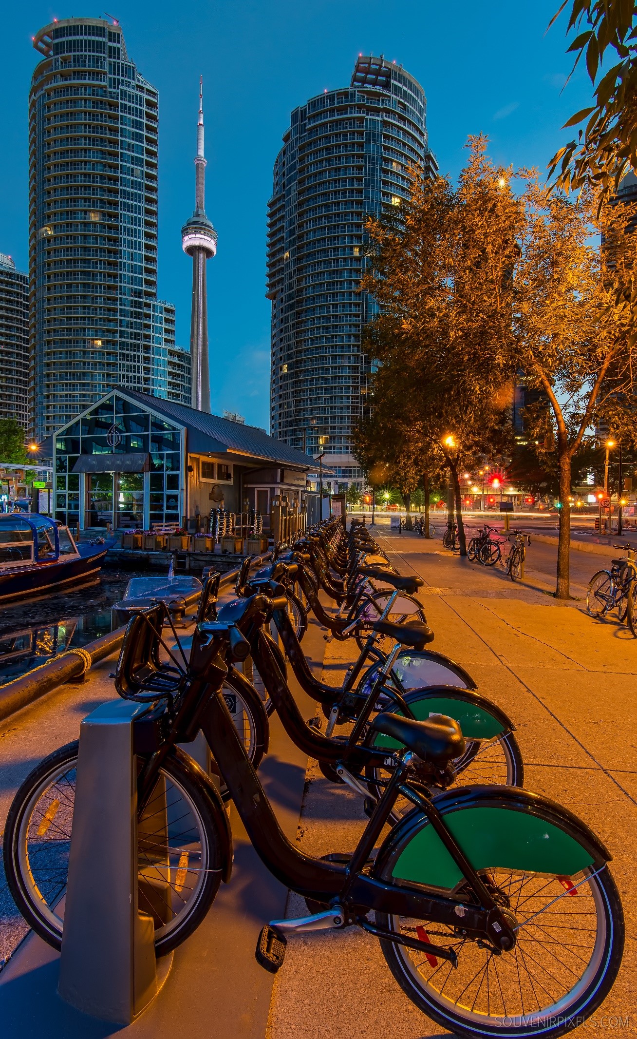
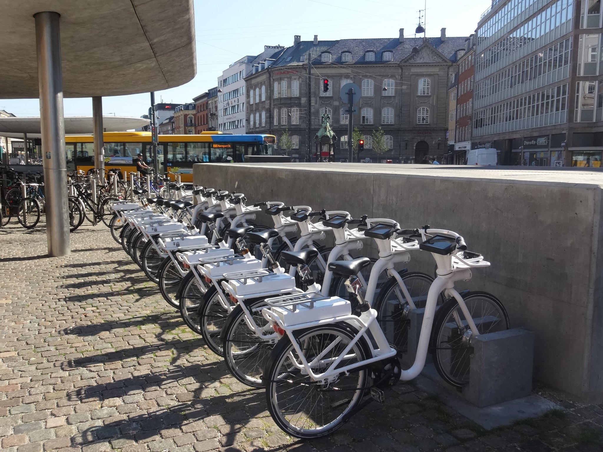
Scientists optimize intranasal anti-depressant drug delivery to the brain

Japanese researchers have succeeded in effective intranasal delivery of modified anti-depressant peptide-based drug to the brain

Drug delivery to the brain has been challenging, owing to disadvantages like systemic absorption, slow axonal transportation, rapid drug degradation, and invasiveness of commonly used techniques. Accordingly, researchers from Japan have successfully attempted to bolster intranasal drug delivery to the brain, making it as effective as other conventional delivery methods, by adding sequences that enhance cell permeability and degradation escape to an anti-depressant drug called glucagon-like peptide 2. Their findings are published in the Journal of Controlled Release.

Clinical researchers working on drug delivery to the brain have been faced with several challenges. Firstly, currently used methods like intracerebroventricular (ICV) administration are extremely invasive. Secondly, other methods like intranasal administration are weighed down by with issues like ineffective cell permeability. Thirdly, even if the drugs gain access into the neurons, their action in the brain is impeded by slow axonal transportation and fast degradation.

After years of meticulous research, a group of Japanese researchers, led by Prof. Chikamasa Yamashita from Tokyo University of Science, working on intranasal drug delivery (IDD) to the brain, have finally achieved success in delivering an anti-depressant drug called glucagon-like peptide 2 (GLP-2) in a mouse model of depression. They modified the drug to accelerate its transportation, while minimizing any degradation, all while achieving the same therapeutic effect as ICV administration, according to their findings published as a research article in the Journal of Controlled Release.

Speaking about the motivation behind pursuing IDD, Prof. Yamashita, the corresponding author of the study, says, “Although there have been more than 20 years of phenomenal IDD research, I had wondered why it has not been put to practical use. Then, I realized that most IDD research had focused on drug delivery through the olfactory epithelium, which accounts for only 2% of human nasal mucosa. Alternatively, my team focused on the central delivery of drugs through the remaining 98% of such mucosa – respiratory epithelium, specifically through the trigeminal nerve.”

The scientists began work with GLP-2, a neuropeptide that has shown therapeutic effects, even in treatment-resistant depression. Given that many drugs entering the body lose their therapeutic effectiveness due to endosomal degradation within the cells, the scientists added a peptide-derived sequence to GLP-2, called a penetration accelerating sequence (PAS) to help evade this degradation. Also, they enhanced the permeability of the drug to the respiratory epithelium, by the addition of another membrane-permeability promoting peptide-derived sequence called as cell penetrating peptides (CPP). Then, they proceeded to test this modified drug in the mouse model of depression.

Their results showed that GLP-2 uptake into the respiratory epithelial cells were enhanced owing to the presence of the CPP. GLP-2 endosomal escape was also enhanced by PAS. In short, this double-modification allowed the effective nose-to-brain delivery of GLP-2. Interestingly, the researchers found that the modified IDDS achieved therapeutic effectiveness in 20 minutes, similar to ICV administration. However, IV administration didn’t demonstrate the intended effect.

Overall, this breakthrough study has taken medicine one step closer to the practical application of intranasal neurological drug administration. Dr. Tomomi Akita, another lead scientist involved in the study, adds, “We hope that our results can be replicated in humans, in the near future. Our study possibly paves the way for futuristic brain delivery applications like nanobiotechnology, and adult genetic engineering, too.”

What a time for neuro-pharmaceutical research, indeed!

Reference

Titles of original papers: Usefulness of cell-penetrating peptides and penetration accelerating sequence for nose-to-brain delivery of glucagon-like peptide-2

Journal: Journal of Controlled Release

DOI: https://doi.org/10.1016/j.jconrel.2021.06.007

Therapeutic Avenue for Inflammatory Diseases

Researchers identify a novel regulatory axis targeting dendritic cell activity and subsequent inflammatory responses in immune disorders

While dendritic cell immunoreceptor (DCIR) is known to mediate inflammation and bone metabolism, ligands that bind DCIR and the mechanisms underlying DCIR activity remain poorly understood. Researchers from Japan have now identified “asialo-biantennary N-glycan”—a glycoprotein present on the surface of bone cells and dendritic cells, as a functional ligand of DCIR. Their findings could help in understanding DCIR’s role in the pathogenesis of autoimmune diseases and bone disorders and develop novel targeted therapies.

Immune cells play a key role in mediating inflammatory responses. Dysregulation in signaling mechanisms that operate across immune cells can trigger chronic inflammatory diseases like rheumatoid arthritis that cause pain and swelling in joints. One such immune cell known to be involved in autoimmune diseases is the dendritic cell. The activity of dendritic cells is regulated by the dendritic cell immunoreceptor (DCIR) present on their surface, which comprises a carbohydrate recognition domain that can bind to sugar moieties present on other proteins or cell surfaces, in a calcium dependent manner. The activity of osteoclast, which is involved in bone degradation, is also regulated by DCIR. However, little is known about the interacting partners of DCIR that help mediate inflammatory responses.

A team of researchers from Japan led by Professor Yoichiro Iwakura of the Department of Experimental Animal Science at the Research Institute for Biomedical Sciences, Tokyo University of Science, have now delved deeper into understanding the mechanisms underlying DCIR activity. In their previous work, the researchers reported that mice deficient in DCIR spontaneously develop arthritis and metabolic bone disorders. Building on this finding, in a recent study published in the Journal of Experimental Medicine, they sought to elucidate DCIR binding partners and immune signaling mechanisms involved in inflammatory diseases. “In this study, we have identified a novel functional ligand of DCIR, likely involved in the pathogenesis of arthritis and other autoimmune diseases like multiple sclerosis. We are hopeful that our work can advance the research of immunology and glycobiology in inflammatory diseases.”, explains Prof. Iwakura.

The researchers began by identifying potential ligands (molecules that bind to cell receptors) of DCIR on immune and bone cells, and found that DCIR binds to glycoproteins present on the surface of macrophages and osteoclasts, the latter differentiating from bone marrow derived macrophages (BMMs), and involved in bone degeneration and remodeling. On further characterization of the glycoprotein, they noted that this interaction was specific to “asialo-biantennary N-glycan (NA2),” a complex carbohydrate moiety comprising various sugar molecules.

Having identified the DCIR ligand, the team next sought to understand the effect of DCIR on osteoclast differentiation and “osteoclastogenesis,” a process contributing to bone loss. Interestingly, cells deficient in DCIR showed a significant increase in the expression of osteoclastogenesis associated genes. As the researchers speculated, expression of DCIR significantly suppressed the differentiation of osteoclasts, thus suggesting DCIR as an independent inhibitor of osteoclastogenesis. Further confirming this finding, a mutant version of DCIR, incapable of recognizing carbohydrate residues, was shown to not exhibit this inhibitory effect.

The role of DCIR in osteoclast differentiation, and its interacting ligand, NA2, now decoded, the team next examined the effect of NA2 on osteoclastogenesis. Consistent with their previous finding, NA2 treatment suppressed the differentiation of osteoclasts from wild-type BMMs but not from cells deficient in DCIR, underscoring the role of NA2 as a specific functional ligand of DCIR that suppresses osteoclastogenesis.

Taking a step further, the researchers treated mouse autoimmune disease models with neuraminidase, an enzyme that removes terminal “sialic acid” residues from N-glycan, thereby enhancing the exposure of NA2. Much to their delight, neuraminidase treatment further suppressed autoimmune diseases like autoimmune arthritis or experimental autoimmune encephalomyelitis, yet again, in a DCIR dependent manner! Furthermore, neuraminidase treatment ameliorated inflammation and associated bone loss in a mouse model of arthritis, thereby confirming their findings in vitro and in vivo. The inhibitory effect of DCIR-NA2 interaction on autoimmune diseases was found to be mediated through suppression of the antigen presenting ability of dendritic cells and subsequent decrease in the activation of other immune cells that contribute to inflammation.

All these findings, together, highlight a novel regulatory mechanism of DCIR signaling involved in the suppression of autoimmunity and excess bone loss. Commenting on the clinical applications of their work, Prof. Iwakura observes, “Our findings are expected to contribute to the understanding of the pathogenesis of human autoimmune diseases such as rheumatoid arthritis and to the development of novel therapies for the treatment of immune and bone metabolic diseases.”

This study indeed takes us a step closer to resolving the complex immune mechanisms of autoimmune diseases, thus paving the way towards effective and targeted treatments.

Novel Light-Based Method Shows the Changing Face of Bioactive Molecules

Using a photocatalyst, scientists have identified the factors behind racemization process of chiral sulfoxides

Chiral sulfoxides, molecules that contain sulfur centers that cannot be superimposed on their mirror images, have wide applications in the chemical industry. This ‘chirality’ of the molecules can make them extremely selective during reactions, which makes them valuable, especially in the pharmaceutical industry. But sulfoxides are famously stable, making their conversion into the desired mirror image challenging. Now, scientists from Tokyo University of Science have developed a new method to convert sulfoxides using light.

The first place you need to look to understand “chirality” are your hands – identical structures that cannot be successfully superimposed over one another. Like our hands, many molecules also exhibit chirality. Different forms of chiral molecules (like the right and left hand) are called enantiomers. Chirality is of particular importance in biology because it endows biological reactions with a high specificity. The reactions that one enantiomer take part in may not necessarily be ones that the other can interact in. This makes enantiomers, and the production of enantiomers, highly desirable to the pharmaceutical industry.

In a solution, enantiomers convert automatically into their mirror-image version by a process called “racemization.” Manipulating the racemization reaction helps produce the desired enantiomer as a product. But some molecules, such as sulfoxides, undergo racemization very slowly. Recently, a team of scientists from Japan—including Professor Hideyo Takahashi, Assistant Professor Kosho Makino, Ms. Kumi Tozawa, and Ms. Yuki Tanaka from Tokyo University of Science—has developed a method to achieve the rapid racemization of chiral sulfoxides, an important group of bioactive compounds and intermediates in chemical reactions. This study has been published in the Journal of Organic Chemistry.

One of the challenges in the racemization of chiral sulfoxides is the requirement of very high temperature (about 200°C) to invert the sulfur-centered pyramid-shaped structure of sulfoxides. But previous research had shown that the racemization of chiral sulfoxides by using light, i.e., photoirradiation, was also possible. This was achieved using a “photosensitizer,” a molecule that donated an electron to the reactants when irradiated with light, thus decreasing the energy required to achieve the reaction. “By conducting the reaction in the presence of a photosensitizer, we were able to achieve the racemization of chiral sulfoxide in much more moderate conditions,” says Prof. Takahashi.

For their work, the team photoirradiated several sulfoxides in the presence of the photosensitizing molecule 2,4,6-triphenylpyrylium tetrafluoroborate (TPT+) and found that effective racemization of a number of different sulfoxides occurred very quickly. Finally, they investigated the kinetics of these reactions and hypothesized a reaction mechanism by which they occurred.

Interestingly, the researchers noticed that not all sulfoxides were racemized by TPT. Certain functional groups such as dimethylamino, pyrrolidyl, and anisyloxy on the sulfoxide hindered the racemization process. To find out why, they assessed the electrochemical potentials (a measure of reactivity) of these functional groups by “cyclic voltammetry.” They saw that these functional groups had lower electrochemical potentials than sulfoxide, implying that they reacted ahead of the sulfoxides, thereby preventing their racemization. “It is our belief that determining electrochemical potentials via cyclic voltammetry will help determine the reactive nature of a photochemical reaction, which will save valuable experimentation and research time,” says Prof. Takahashi.

The study emerges as a distinguished addition to research on manipulating the chirality of molecules. As per Prof. Takahashi, “Our photosensitization-based rapid racemization technology can be used for dynamic asymmetric-induced reactions for obtaining the desired enantiomers.”

Indeed, such research is sure to advance the field of chiral active pharmaceuticals and functional materials!

Reference
Titles of original papers: Rapid photoracemization of chiral alkyl aryl sulfoxides
Journal: Journal of Organic Chemistry
DOI: https://doi.org//10.1021/acs.joc.1c02320

Plumbing the Depths: Defect Distribution in Ion-Implanted SiC Diodes

Researchers reveal that aluminum implantation doping in p-type bipolar semiconductors creates defects many layers deeper than the implantation site

Introducing a vertical arrangement of n and p layers into the drift layer of semiconductors to enable bipolar operation is a way around the ‘unipolar limit’ problem in semiconductors. But defect generation during the fabrication of such devices is a matter of concern. Researchers have examined the depth and distribution of defects formed by aluminum ion implantation in silicon carbide bipolar diodes to identify ways to induce efficient conductivity modulation.

Silicon carbide (SiC) unipolar semiconductors are in wide commercial use, but their operations are limited by a trade-off relationship between breakdown voltage and specific resistance of the drift layer, or specific on-resistance. Including a super junction structure, which refers to an arrangement of n and p layers in trenches in the drift layer, or enabling bipolar operation in the device, provides a way to overcome this unipolar limit. Bipolar operation brings about a large decrease in on-resistance by inducing a conductivity modulation in the drift layer. But bipolar operation is not without its disadvantages. Conduction and switching losses in bipolar devices need to be carefully balanced.

P-type contact layers in semiconductors are generally formed via aluminum (Al) doping. Al doping can be achieved in two ways – epitaxial or ion implantation. Epitaxial growth involves the layer by layer deposition of semiconductor materials on a substrate, whereas ion implantation entails bombarding the semiconductor layers with high energy charged particles. But ion implantation leads to the formation of defects deep in the semiconductor layers, which could have a critical effect on conductivity modulation.

In a recent study published in Physica Status Solidi (b), researchers from Japan investigated the depth distribution of defects in SiC bipolar diodes that were formed by Al doping. “Our findings will help with the optimum design of SiC power devices, which will soon be employed in electric vehicles, trains etc. These results will ultimately help improve the performance, as well as the size and energy consumption of traction systems in vehicles and trains,” says Associate Professor Dr. Masashi Kato of Nagoya Institute of Technology, who led the study.

To study the depth distribution of defects, the research team fabricated two SiC PiN diodes with Al doped p-layers, one through epitaxial growth and the other through ion implantation. They then studied the distribution of defects in both diodes using conventional ‘deep level transient spectroscopy’ (DLTS) and characterized its properties using cathodoluminescence (CL). They found that p-type layer deposition by epitaxial growth did not cause damage in the adjacent n-type layers, but that the growth showed slight instability that led to the formation of deep level defects. The specific on-resistance of this diode was also low, thanks to the effects of conductivity modulation.

For the diode formed by ion implantation, however, the researchers found that Al doping achieved a high specific on-resistance without influencing conductivity modulation. Moreover, the researchers observed that the defects in the semiconductor device penetrated to a minimum of 20 µm from the implantation region. “Our study shows that the ion implantation in SiC bipolar devices need to be processed at least 20 µm away from the active regions,” explains Dr. Kato.

The low power consumption of SiC power devices mean that they will be essential in the future as climate change increases and the fossil fuel energy crisis worsens. Improving semiconductor technology rapidly so that it can take its rightful place on the world stage is of paramount importance. With strong results like this to inform future research and manufacturing, we may realize this future sooner than expected!

New Tool Can Detect a Precursor of Engine-Destroying Combustion Instability

Scientists develop a tool that uses machine learning and a dynamical systems-based approach to identify a precursor of combustion instability in engines

Combustion engines, like those in aircrafts, remain at risk of fatal damage by a phenomenon called “combustion oscillations,” where pressure fluctuations inside the engine become large. Now, researchers from Japan have developed a novel tool to detect a precursor to combustion oscillations using machine learning and a dynamical systems-based approach, opening doors to the prediction and prevention of related fatal damage to engines.

Combustion engines have been around since the late 18th century, although they did not gain popularity until over 50 years later. Now, they are practically ubiquitous, powering anything from cars and airplanes to turbines.

The part of the combustion engine in which the fuel is burnt (in the presence of oxygen) is called the combustor. The lifespan of a combustor can be limited by a phenomenon called “thermoacoustic combustion oscillations.” When thermoacoustic oscillations become too large or out of control, it causes fatal damage to combustors, which can have enormous financial and human consequences.

Detecting combustion oscillations and preventing damage is a key effort in the field of thermal engineering. Recently, a team of scientists from Japan—including Hiroshi Gotoda, Yuhei Shinichi, and Naohiro Takeda from Tokyo University of Science, as well as Seiji Yoshida and Takeshi Shoji from the Japan Aerospace Exploration Agency (JAXA)—have developed a promising tool for the detection of a precursor of thermoacoustic oscillations. The study was made available online on May 10, 2021 and published in volume 59 of the American Institute of Aeronautics and Astronautics Journal on October 1, 2021.

“In our study, we have shown that the methodology combining dynamical systems theory and machine learning can be useful for detecting predictive combustion oscillations in multisector combustors, such as those in aircraft engines,” says Prof. Gotoda, who headed the study.

The team conducted combustion experiments with varying fuel flow rates in a staged multisector combustor developed by JAXA.

The scientists used the data from these experiments to train a machine learning algorithm called ‘support vector machine (SVM).’ The SVM allowed them to classify the combustion into three states—stable, transitional, and combustion oscillations. The pressure fluctuations in the transitional state are key to predicting future combustion oscillations. In the transitional state, pressure fluctuations transition from being small-amplitude and aperiodic to being large-amplitude and periodic. Amplitude represents the ‘largeness’ of the fluctuation, whereas periodicity describes the repetition of the fluctuation.

“The findings of this study will contribute greatly towards developing a method to detect combustion oscillations in advance in aircraft engines,” reveals Prof. Gotoda.

These findings could have far-reaching consequences, paving the way for confident and timely predictions of combustion oscillations, with the potential to save billions of dollars and human lives.

New treatment for inflammatory bowel disease

In a recent study published in Frontiers in Immunology, Prof. Chiharu Nishiyama, Kazuki Nagata, and Ayumi Okuzumi from the Tokyo University of Science and Prof. Hiroshi Nagase from the University of Tsukuba attempted to understand the effects of opioids on the immune system. They tested the effects of KNT-127—an artificially synthesized opioid that activates delta-opioid receptors—on immune responses in live animal and cell culture experiments.

When mice with inflammatory bowel disease (IBD) were treated with KNT-127, they showed a reduction in the severity of colitis—a form of colon inflammation—indicated by lower weight loss and colon atrophy and improved disease activity scores. Similar results were also obtained in a recovery model, confirming the beneficial effects of KNT-127 against colonic inflammation.

Although these results were promising, an important caveat still loomed. “Before proceeding with additional experiments, we had to rule out the role of CNS opioid receptors in the anti-inflammatory effects of KNT-127,” says Prof. Nishiyama, the lead researcher on the study.

To address this, the researchers performed similar experiments with YNT-2715, a peripheral KNT-127 that cannot cross over from the blood to the brain. The results were similar to those observed with KNT-127, confirming that its anti-inflammatory effects were indeed CNS-independent.

Encouraged by this, the group examined other immune-related effects of KNT-127 treatment in the colitis model. They found that during disease progression, the opioid reduced the serum levels of IL-6, a pro-inflammatory factor, while also decreasing the number of macrophages in the mesenteric lymph nodes (MLNs).

Interestingly, they also observed an increase in the number of regulatory T cells (Tregs) in MLNs. Together, their results showed that KNT-127 suppresses the inflammation caused by macrophages during disease progression and enhances the anti-inflammatory response due to Tregs during recovery.

Finally, to understand the direct effects of KNT-127 on immune cells, the researchers performed in vitro experiments in which they treated macrophages derived from bone marrow or T cells from the spleen with the drug. The results were consistent with those from animal experiments, revealing increased secretion of the pro-inflammatory signals as well as enhanced development of Tregs in response to KNT-127 treatment.

Altogether, the findings demonstrated that KNT-127 can directly act on immune cells and reduce the severity of inflammation, making it a good candidate for the treatment of IBD.

“Several people around the world suffer from diseases related to colon inflammation, and so far, optimal treatment strategies are lacking. Our findings show that KNT-127 and other activators of opioid receptors could be promising therapeutic options for such diseases,” comments Prof. Nagase, the chief drug developer behind the synthetic opioid, while also cautioning of the road ahead.

“Of course, before these drugs are used clinically, additional experiments will be required to elucidate how they exert their immunomodulatory functions and what their effects on other immune diseases are,” he adds.

Nevertheless, Prof. Nishiyama and her team are confident that their study represents an important milestone, not only towards the treatment of IBD but also towards our understanding of the “brain-gut axis”—the interrelationship between brain and gut function—which has received increasing attention in recent years.

“Today, we know that poor mental health has physical manifestations. For example, stress worsens inflammation in the gut, which in turn affects the health of the brain. Our results on the immune-related effects of opioids, which commonly act on the brain, is a step toward unravelling the biological mechanisms that govern the reciprocative relationship of gut health and the immune system with the CNS,” mentions Prof. Nishiyama, excited about what the future holds.

Hidden in the Seeds: Bacteria found to survive the harsh interior of Passion Fruit seeds

For the first time ever, a research group consisting of Dr. Toshiki Furuya, Associate Professor at the Department of Applied Biological Science, Tokyo University of Science, Japan, has succeeded in isolating bacteria from the seeds of passion fruit (Passiflora edulis). Their research, which also unearths the surviving mechanisms of these bacteria inside the seeds, has been published in the journal MicrobiologyOpen.

In their study, the scientists focused on the seeds of P. edulis. The seeds of this fruit are full of secondary metabolites with strong antimicrobial properties, such as resveratrol and piceatannol—the latter present at high levels of up to 2.2 mg/g.

As Dr. Furuya reveals the rationale behind choosing passion fruit seeds for the study, “The extraordinarily high concentration of piceatannol protects P. edulis seeds from microorganisms. We thought it would be interesting to know if any endophytic microorganism could survive this extreme environment and if yes, how.”

Earlier reports showed that endophytes capable of surviving in an environment rich in biologically active compounds possessed biocatalytic activities related to the metabolism of these compounds. The fact that their biocatalytic potential could be exploited for therapeutic purposes made the scientists even more eager to explore the presence of endophytic bacteria.

The scientists collected and surface-sterilized the seeds of naturally grown P. edulis before either cutting or crushing them and placing them on solid agar-based growth media to check for microbial growth. While no microbial colony appeared from the cut or homogenized seeds, interestingly, the seedlings sprouting from the cut seeds, when exposed to growth media, gave rise to microbial colonies. The scientists then performed sequencing to identify the bacteria that appeared on the agar plate.

The findings were remarkable. From the seedlings, the scientists isolated 19 strains, including three previously unreported strains of bacteria from various genera. They hypothesized that inside the seeds, piceatannol exerted bacteriostatic (or “bacterial growth-stalling”) rather than bactericidal (or “bacteria-killing”) effects on the residing bacteria.

Ms. Aoi Ishida, the co-author of the study explains: “Due to the presence of a high concentration of piceatannol, the growth of the bacteria was stagnated inside the seed, but when transmitted to the next-generation seedlings during germination, the bacteria were relieved from the effect of piceatannol and able to grow again.”

The scientists also found one of the bacteria, Brevibacterium sp. PE28-2, to possess the ability to convert resveratrol and piceatannol to their respective derivatives. This is the first endophyte shown to exhibit such activity.

Dr. Furuya and Ms. Ishida are very hopeful that the method established in this study is expected to be effective in isolating several useful endophytic bacteria from a variety of plants. Moreover, considering the current focus on engineering new biomolecules with diverse applications, the results of this study would accelerate research on seed endophytic bacteria.

“Second-Hand” psychological stress can lead to depression in mice, finds study

In a pioneering study, scientists from Japan established that psychological stress can negatively affect neurogenesis and cause depression in a mouse model.

Recent research has explained how vicarious social defeat can cause psychological stress in mice. This involves the mouse being made to experience the defeat of another mouse in an experimental social setting. Using this model, a group of scientists from Japan attempted to establish a link between depressive symptoms and hippocampal neurogenesis.

Professor Akiyoshi Saitoh from Tokyo University of Science, one of the lead authors of the study, further explains the motivation behind this research, “The number of individuals suffering from depression has been on the rise the world over. However, the detailed pathophysiology of depression still remains to be elucidated. So, we decided to focus on the possible mechanism of psychological stress in adult hippocampal neurogenesis, to understand its role in depressive disorders.” The study was published in the journal Behavioural Brain Research.

After exposing the mice to chronic vicarious social defeat stress, Prof. Saitoh and the team, including Mr Toshinori Yoshioka and Dr Daisuke Yamada from Tokyo University of Science, analyzed their behaviour and brains in close detail.

Aside from behavioural deficits like social withdrawal, the stressed mice also showed a significant decrease in the survival rate of newborn neurons in the dentate gyrus, a region in the hippocampus responsible for sensory perception and memory, compared to the non-stressed controls. This condition persisted for up to four weeks, after “stressing” the mice. However, cell growth, differentiation, and maturation did not differ between the groups of mice during the period of observation. Notably, the cell survival rate was restored in the stressed mice after treatment with a chronic antidepressant called fluoxetine.

Regarding the results, Mr. Toshinori Yoshioka adds, “We have found out that chronic mental stress affects the neurogenesis of the hippocampal dentate gyrus. Also, we believe that this animal model will play an important role in elucidating the pathophysiology of depression, and in the development of the corresponding novel drug.”

Overall, this study has provided important insights into the pathophysiology of depression. Also, it goes without saying how this study paves the way for future research into the role of psychological stress in depression.

Scientists unravel the mysteries of irreversibility in electrochromic thin films

Scientists from Japan have quantitatively evaluated ion-trapping-induced degradation in lithium intercalated tungsten oxide films

In a recent study published in Applied Surface Science (made available online on August 13 2021, and to be published in Volume 568 of the journal on December 1 2021), scientists from the Tokyo University of Science and the National Institute for Materials Science (NIMS), Japan, collaborated to quantitatively assess the irreversibility of LixWO3 thin films.

Discussing the key concerns that the study addresses, Associate Professor Tohru Higuchi from Tokyo University of Science, who led the study, observes “There are two critical questions that arise: First, is irreversible Li2WO4 formation different from irreversible Li+ trapping? Second, can these irreversible components coexist?”

He adds, “Conventional measures are unable to differentiate between the two irreversible components. As a result, we conducted a quantitative examination to offer solid answers to these questions.”

The scientists devised a quantitative evaluation method that combines in situ hard X-ray photoelectron spectroscopy (HAXPES) and electrochemical measurements. HAXPES is used to investigate buried interfaces, whereas electrochemical tests are used to examine corrosion properties. The intercalation of Li+ results in a redox reaction that changes the oxidation state of tungsten (W) ions from W6+ to W5+.

Based on this change, HAXPES can evaluate “reversible Li+” and “irreversible Li+ trapping.” However, evaluating “irreversible Li2WO4 formation” using HAXPES is challenging.

Dr Takashi Tsuchiya, a Principal Researcher at NIMS and co-author of the study, explains why: “W ions in Li2WO4 have a stable oxidation state because they exist in the W6+ form. As a result, HAXPES is unable to evaluate the irreversibility caused by Li2WO4 formation. Electrochemical measurements, on the contrary, can distinguish ‘reversible Li+’ from the two irreversible components. Therefore, integrating both methods enables the distinction and quantitative evaluation of all three components.”

To conduct the electrochemical measurements, the scientists built a LixWO3-based redox transistor on the flat surface of a lithium-ion conducting glass ceramic (LICGC). They also built an electrochemical cell with a WO3 thin film as the semiconductor and a LICGC substrate as the electrolyte to conduct HAXPES measurements.

Furthermore, they employed in situ Raman spectroscopy to assess the influence of Li+ insertion on the LixWO3 structure. They were able to successfully determine the increase in crystallinity caused by Li+ insertion. The proportions of reversible Li+, irreversible Li2WO4 formation, and irreversible Li+ trapping were calculated to be 41.4%, 50.9%, and 7.7%, respectively.

The scientists believe that their study will help develop and design improved EC materials and devices.

“For several years, the main impetus for EC research and development has been potential applications in energy-efficient buildings and aircraft. However, there are several other applications as well, such as the energy-saving and vision-friendly electronic paper displays,” says Dr Kazuya Terabe, Principal Investigator of the International Center for Materials Nanoarchitectonics at NIMS and a co-author of the study.

“Moreover, our findings broaden the application possibilities by providing the basis for the future development of high-performance WO3-based EC devices.”

Untangling the irreversibility conundrum is certainly a big step forward, but there is still much work to be done, although the pace is sure to go up.

Self-powered diaper sensors that monitor urine sugar levels

Monitoring urine sugar levels is important during the early stages of diabetes, and diaper sensors represent an attractive solution. In a recent study published in ACS Sensors, Associate Professor Isao Shitanda, Professor Masayuku Itagaki, and Mr Yuki Fujimura from Tokyo University of Science (TUS), Japan, present a promising approach to realizing self-powered diaper sensors that can generate energy directly from urine.

This work was done in collaboration with Associate Professor Seiya Tsujimura from the University of Tsukuba, Japan. Worth noting, this work is in line with other research efforts of Dr. Shitanda and his colleagues to develop self-powered biosensors like a lactate sensor energized entirely by sweat.

The scientists developed a paper-based biofuel cell that, through a pair of reduction-oxidation reactions, outputs electrical power proportional to the amount of glucose in the urine. Important considerations in the design of such biofuel cells are the amount of urine needed to generate enough power and the overall stability and durability of the device.

With this in mind, the scientists developed a special anode, the negative terminal of an electrochemical cell, using a process known as “graft polymerization” that allowed them to firmly anchor glucose-reactive enzymes and mediator molecules to a porous carbon layer, which served as the base conductive material.

The scientists tested their self-powered biosensor in diapers using artificial urine at various glucose concentrations. They used the generated energy to power up a Bluetooth Low Energy transmitter, and remotely monitored the measured concentration using a smartphone. They found that the biofuel cell could detect urine sugar in a very short time (within 1 second).

“Besides monitoring glucose in the context of diabetes, diaper sensors can be used to remotely check for the presence of urine if you stock up on sugar as fuel in advance. In hospitals or nursing care sites, where potentially hundreds of diapers have to be checked periodically, the proposed device could take a great weight off the shoulders of caregivers,” comments Dr Shitanda.

In short, the sensor that Dr Shitanda’s team has engineered can not only prevent diabetes but also make diaper management more efficient and responsive without compromising the environment. “We believe the concept developed in this study could become a very promising tool towards the general development of self-powered wearable biosensors,” says Dr Shitanda.