


Wearable sensors are becoming a promising tool in personalized healthcare and exercise monitoring. In a recent study, researchers from Japan develop a novel wearable chemical sensor capable of measuring the concentration of chloride ions in sweat. By using a heat-transfer printing technique, the proposed sensor can be applied to the outer surface of common textiles to prevent skin irritation and allergies, and could also be useful in the early detection of heat stroke and dehydration.
The remarkable level of miniaturization possible in modern electronics has paved the way for realizing healthcare devices previously confined to the realm of science fiction. Wearable sensors are a prominent example of this. As the name suggests, these devices are worn on the body, usually directly on the skin. They can monitor important bodily parameters, including heart rate, blood pressure, and muscle activity.
Some wearable sensors can also detect chemicals in bodily fluids. For instance, sweat biosensors can measure the concentration of ions in sweat, providing information on their levels in blood. However, designing such chemical sensors is more complex than physical sensors. Direct contact between a wearable chemical sensor and skin can cause irritation and allergies. In contrast, if the sensor is fabricated directly on a wearable textile, its accuracy decreases due to surface irregularities.
In a recent study, a research team, led by Associate Professor Isao Shitanda of the Tokyo University of Science (TUS) in Japan, has developed an innovative sweat biosensor that addresses the aforementioned problems. Their work, published online in ACS Sensors on June 15, 2023, describes the use of a technique called “heat-transfer printing” to fix a thin, flexible chloride ion sensor onto a textile substrate. The study was co-authored by Dr. Masahiro Motosuke, Dr. Tatsunori Suzuki, Dr. Shinya Yanagita, and Dr. Takahiro Mukaimoto of TUS.
“The proposed sensor can be transferred to fiber substrates, and thus can be incorporated into textiles such as T-shirts, wristbands, and insoles,” explains Dr. Shitanda. “Further, health indicators such as chloride ion concentration in sweat can be measured by simply wearing them.”
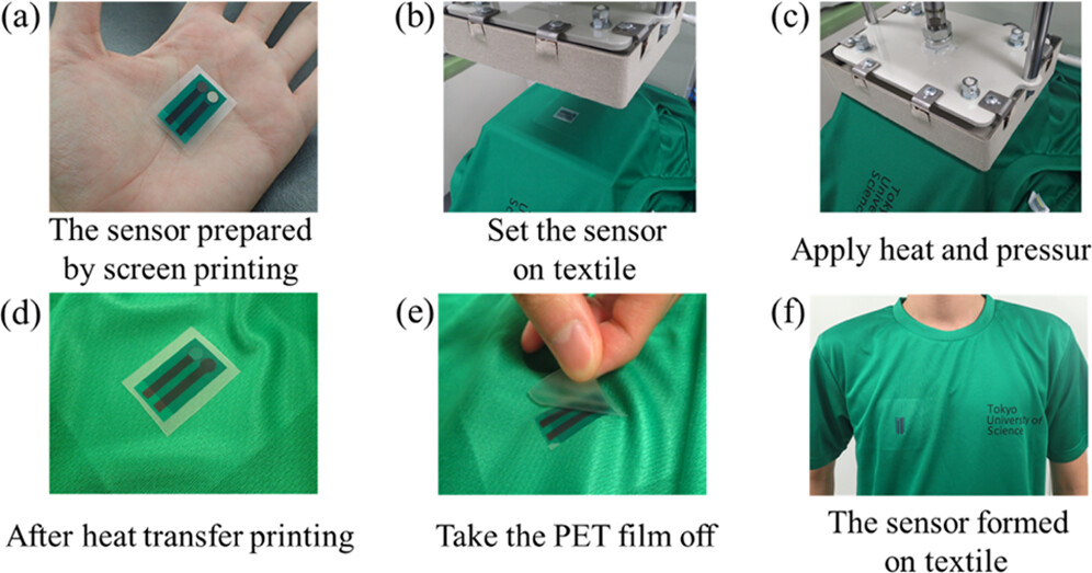
The heat-transfer printing approach offers several advantages. For one thing, the sensor is transferred outside of the piece of clothing, which prevents skin irritation. In addition, the wicking effect of the textile helps spread the sweat evenly between the electrodes of the sensor, creating a stable electrical contact. Moreover, printing the sensor on a flat surface and then transferring it prevents the formation of blurred edges that commonly occur when printing directly onto a textile.
The researchers carefully selected the materials and electrochemical mechanisms of the sensor to avoid risking an allergic reaction for the wearer. After developing the sensor, they conducted various experiments using artificial sweat to verify its accuracy in measuring chloride ion concentration. The change in the electromotive force of the sensor was −59.5 mTV/log CCl−. Additionally, it displayed a Nernst response and a linear relationship with the concentration range of chloride ions in human sweat. Moreover, no other ions or substances typically present in sweat were found to interfere with the measurements.
Lastly, the team tested the sensor on a volunteer who exercised on a static bicycle for 30 minutes, by measuring their perspiration rate, chloride ion levels in blood, and saliva osmolality every five minutes to compare with the data previously gathered by the sensor. The proposed wearable sensor could reliably measure the concentration of chloride ions in sweat.
The sensor can also transmit data wirelessly, making it useful for real-time health monitoring. “Since chloride is the most abundant electrolyte in human sweat, measuring its concentration provides an excellent indicator of the body’s electrolyte balance and a useful tool for the diagnosis and prevention of heat stroke,” remarks Dr. Shitanda.
This research thus demonstrates the potential of using wearable ion sensors for the real-time monitoring of sweat biomarkers, facilitating personalized healthcare development and athlete training management.


.jpg)
.png)