INHA University
Published on 7 September 2026




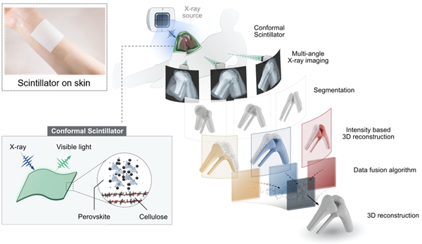
▲ Overview of the film-based X-ray imaging material capable of conforming to curved surfaces of the human body and the AI-based 3D reconstruction system.

▲Realization of 3D images through the reconstruction of segmented X-ray images using AI.
Professor Donghyuk Park’s research team from the Department of Chemical Engineering at Inha University recently developed a 3D X-ray image reconstruction technology using traditional Korean Hanji through a joint research project with domestic and international researchers.
This technology reconstructs internal structures of the human body into 3D images using only three X-ray images. Traditionally, obtaining 3D images using X-rays requires dozens to hundreds of images, as in computed tomography (CT). However, the research team demonstrated the potential to reconstruct 3D structures using only a small number of X-ray images by utilizing artificial intelligence (AI).
The research team also developed a thin and flexible X-ray detection film based on cellulose, the main component of traditional Hanji.
An X-ray detector is a device that detects X-rays and converts them into images. Unlike conventional rigid flat-panel detectors, the film developed in this study can bend like skin and conform closely to curved surfaces. This can help reduce image distortion that may occur during the imaging process.
The key technology involves directly growing crystals on the surface of cellulose. The research team grew lead-free, environmentally friendly perovskite crystals on cellulose fibers. Perovskites are materials used to convert X-rays into light. Through this approach, the research team was able to maintain the film’s X-ray-to-light conversion performance while reducing its thickness.
The team then combined the technology with AI-based image reconstruction. The AI was designed to automatically identify specific areas of interest, such as bones, from three X-ray images taken from different angles. The identified information was then combined to reconstruct a single three-dimensional model.
The accuracy of the technology was validated through experiments using actual bird leg bones. The results demonstrated the potential to obtain 3D diagnostic information using existing X-ray imaging infrastructure without the need for expensive CT equipment.
This technology is significant because it has the potential to reduce radiation exposure and equipment costs. It is expected to have applications in emergency medical settings and regions with limited medical infrastructure in the future.
The study was recently published in the prestigious international academic journal ‘Nature Communications’ in recognition of presenting new possibilities for 3D X-ray image reconstruction by combining low-cost, eco-friendly materials with AI technology.
The research was jointly conducted by the research teams of Professor Donghyuk Park of the Department of Chemical Engineering at Inha University, Professor Kyusang Lee of the University of Virginia (UVA) in the United States, Professor Sang-eun Jo of Dongguk University, and Professor Kyunghak Kim of Hanyang University. Dr. Seokho Kim, a co-first author, earned his Ph.D. in Chemical Engineering from Inha University and is currently working as a postdoctoral researcher at the University of Virginia. He led the research together with Dr. Byungjun Bae and Dr. Dowan Kim.
Professor Donghyuk Park of the Department of Chemical Engineering said, “This research is significant in that it presents new possibilities for X-ray imaging diagnosis by combining an environmentally friendly material based on traditional Korean Hanji with artificial intelligence technology. We will continue follow-up research to develop this technology into a low-cost, low-dose 3D X-ray imaging technology.”

▲(From left) Dr. Seokho Kim (UVA), Dr. Byungjun Bae (UVA), Dr. Dowan Kim (UVA), Professor Kyusang Lee (UVA), and Professor Donghyuk Park (Inha University)